3
views
0
recommends
+1 Recommend
0 collections
    0
    shares
      • Record: found
      • Abstract: found
      • Article: found
      Is Open Access

      Metal Ion Binding to the Amyloid β Monomer Studied by Native Top-Down FTICR Mass Spectrometry

      research-article

      Read this article at

      Bookmark
          There is no author summary for this article yet. Authors can add summaries to their articles on ScienceOpen to make them more accessible to a non-specialist audience.

          Abstract

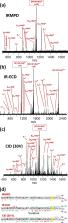
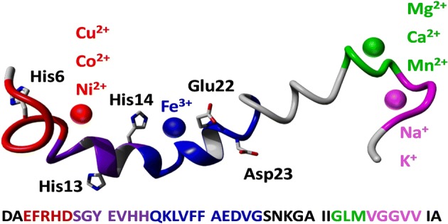
          Native top-down mass spectrometry is a fast, robust biophysical technique that can provide molecular-scale information on the interaction between proteins or peptides and ligands, including metal cations. Here we have analyzed complexes of the full-length amyloid β (1-42) monomer with a range of (patho)physiologically relevant metal cations using native Fourier transform ion cyclotron resonance mass spectrometry and three different fragmentation methods—collision-induced dissociation, electron capture dissociation, and infrared multiphoton dissociation—all yielding consistent results. Amyloid β is of particular interest as its oligomerization and aggregation are major events in the etiology of Alzheimer’s disease, and it is known that interactions between the peptide and bioavailable metal cations have the potential to significantly damage neurons. Those metals which exhibited the strongest binding to the peptide (Cu 2+, Co 2+, Ni 2+) all shared a very similar binding region containing two of the histidine residues near the N-terminus (His6, His13). Notably, Fe 3+ bound to the peptide only when stabilized toward hydrolysis, aggregation, and precipitation by a chelating ligand, binding in the region between Ser8 and Gly25. We also identified two additional binding regions near the flexible, hydrophobic C-terminus, where other metals (Mg 2+, Ca 2+, Mn 2+, Na +, and K +) bound more weakly—one centered on Leu34, and one on Gly38. Unexpectedly, collisional activation of the complex formed between the peptide and [Co III(NH 3) 6] 3+ induced gas-phase reduction of the metal to Co II, allowing the peptide to fragment via radical-based dissociation pathways. This work demonstrates how native mass spectrometry can provide new insights into the interactions between amyloid β and metal cations.

          Electronic supplementary material

          The online version of this article (10.1007/s13361-019-02283-7) contains supplementary material, which is available to authorized users.

          Related collections

          Most cited references88

          • Record: found
          • Abstract: found
          • Article: not found

          Studying noncovalent protein complexes by electrospray ionization mass spectrometry.

          J A Loo (2015)
          Electrospray ionization mass spectrometry has been used to study protein interactions driven by noncovalent forces. The gentleness of the electrospray ionization process allows intact protein complexes to be directly detected by mass spectrometry. Evidence from the growing body of literature suggests that the ESI-MS observations for these weakly bound systems reflect, to some extent, the nature of the interaction found in the condensed phase. Stoichiometry of the complex can be easily obtained from the resulting mass spectrum because the molecular weight of the complex is directly measured. For the study of protein interactions, ESI-MS is complementary to other biophysical methods, such as NMR and analytical ultracentrifugation. However, mass spectrometry offers advantages in speed and sensitivity. The experimental variables that play a role in the outcome of ESI-MS studies of noncovalently bound complexes are reviewed. Several applications of ESI-MS are discussed, including protein interactions with metal ions and nucleic acids and subunit protein structures (quaternary structure).
            Bookmark
            • Record: found
            • Abstract: found
            • Article: not found

            Metal binding and oxidation of amyloid-beta within isolated senile plaque cores: Raman microscopic evidence.

            Alzheimer's disease (AD) is characterized by the deposition of amyloid plaques in the parenchyma and vasculature of the brain. Although previous analytical studies have provided much information about the composition and structure of synthetic amyloid-beta fibrils, there is, surprisingly, a dearth of data on intact amyloid plaques from AD brain. Therefore, to elucidate the structure and detailed composition of isolated amyloid plaque cores, we utilized a high-resolution, nondestructive technique, Raman microscopy. The data are of very high quality and contain detailed information about protein composition and conformation, about post-translational modification, and about the chemistry of metal binding sites. Remarkably, spectra obtained for senile plaque (SP) cores isolated from AD brain are essentially identical both within and among brains. The Raman data show for the first time that the SP cores are composed largely of amyloid-beta and confirm inferences from X-ray studies that the structure is beta-sheet with the additional possibility that this may be present as a parallel beta-helix. Raman bands characteristic of methionine sulfoxide show that extensive methionine oxidation has occurred in the intact plaques. The Raman spectra also demonstrate that Zn(II) and Cu(II) are coordinated to histidine residues in the SP cores, at the side chains' N(tau) and N(pi) atoms, respectively. Treatment of the senile plaques with the chelator ethylenediaminetetraacetate reverses Cu binding to SP histidines and leads to a broadening of amide features, indicating a "loosening" of the beta-structure. Our results indicate that Abeta in vivo is a metalloprotein, and the loosening of the structure following chelation treatment suggests a possible means for the solubilization of amyloid deposits. The results also reveal a direct chemical basis for oxidative damage caused by amyloid-beta protein in AD.
              Bookmark
              • Record: found
              • Abstract: found
              • Article: not found

              Iron accumulation in Alzheimer disease is a source of redox-generated free radicals.

              Damage from free radicals has been demonstrated in susceptible neuronal populations in cases of Alzheimer disease. In this study, we investigated whether iron, a potent source of the highly reactive hydroxyl radical that is generated by the Fenton reaction with H2O2, might contribute to the source of radicals in Alzheimer disease. We found, using a modified histochemical technique that relies on the formation of mixed valence iron complexes, that redox-active iron is associated with the senile plaques and neurofibrillary tangles-the pathological hallmark lesions of this disease. This lesion-associated iron is able to participate in in situ oxidation and readily catalyzes an H2O2-dependent oxidation. Furthermore, removal of iron was completely effected using deferoxamine, after which iron could be rebound to the lesions. Characterization of the iron-binding site suggests that binding is dependent on available histidine residues and on protein conformation. Taken together, these findings indicate that iron accumulation could be an important contributor toward the oxidative damage of Alzheimer disease.
                Bookmark

                Author and article information

                Contributors
                f.lermyte@warwick.ac.uk
                Journal
                J Am Soc Mass Spectrom
                J. Am. Soc. Mass Spectrom
                Journal of the American Society for Mass Spectrometry
                Springer US (New York )
                1044-0305
                1879-1123
                26 July 2019
                26 July 2019
                2019
                : 30
                : 10
                : 2123-2134
                Affiliations
                [1 ]GRID grid.7372.1, ISNI 0000 0000 8809 1613, School of Engineering, , University of Warwick, ; Coventry, CV4 7AL UK
                [2 ]GRID grid.7372.1, ISNI 0000 0000 8809 1613, Department of Chemistry, , University of Warwick, ; Coventry, CV4 7AL UK
                [3 ]GRID grid.9757.c, ISNI 0000 0004 0415 6205, Institute for Science and Technology in Medicine, , Keele University, ; Stoke-on-Trent, Staffordshire ST4 7QB UK
                Author information
                http://orcid.org/0000-0001-7371-4475
                http://orcid.org/0000-0002-1864-9700
                http://orcid.org/0000-0001-5486-091X
                http://orcid.org/0000-0002-3647-2611
                http://orcid.org/0000-0002-4035-0369
                http://orcid.org/0000-0002-6474-5357
                http://orcid.org/0000-0002-2683-5546
                http://orcid.org/0000-0001-9160-1941
                http://orcid.org/0000-0002-6588-6274
                http://orcid.org/0000-0002-8423-4183
                Article
                2283
                10.1007/s13361-019-02283-7
                6805827
                31350722
                0acbbfcb-c348-46be-9783-9593784ea6cb
                © The Author(s) 2019

                Open Access This article is distributed under the terms of the Creative Commons Attribution 4.0 International License ( http://creativecommons.org/licenses/by/4.0/), which permits unrestricted use, distribution, and reproduction in any medium, provided you give appropriate credit to the original author(s) and the source, provide a link to the Creative Commons license, and indicate if changes were made.

                History
                : 10 June 2019
                : 1 July 2019
                : 1 July 2019
                Funding
                Funded by: Engineering and Physical Sciences Research Council
                Award ID: EP/N033191/1
                Award ID: EP/N033140/1
                Award Recipient :
                Categories
                Research Article
                Custom metadata
                © American Society for Mass Spectrometry 2019

                Analytical chemistry
                native mass spectrometry,amyloid beta,peptide-metal complex,native top-down,mass spectrometry,electron capture dissociation,collision-induced dissociation,infrared multiphoton dissociation,radical-directed dissociation

                Comments

                Comment on this article