2
views
0
recommends
+1 Recommend
0 collections
    0
    shares
      • Record: found
      • Abstract: found
      • Article: found
      Is Open Access

      Inflammasome Activation Induced by a Snake Venom Lys49-Phospholipase A 2 Homologue

      research-article

      Read this article at

      Bookmark
          There is no author summary for this article yet. Authors can add summaries to their articles on ScienceOpen to make them more accessible to a non-specialist audience.

          Abstract

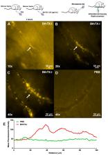
          Background: Snake venom phospholipases A 2 (PLA 2s) have hemolytic, anticoagulant, myotoxic, oedematogenic, bactericidal, and inflammatory actions. BthTX-I, a Lys49-PLA 2 isolated from Bothrops jararacussu venom, is an example of Lys49-PLA 2 that presents such actions. NLRP3 is a cytosolic receptor from the NLR family responsible for inflammasome activation via caspase-1 activation and IL-1β liberation. The study of NLRs that recognize tissue damage and activate the inflammasome is relevant in envenomation. Methods: Male mice (18–20 g) received an intramuscular injection of BthTX-I or sterile saline. The serum was collected for creatine-kinase (CK), lactate dehydrogenase (LDH), and interleukin-1β (IL-1β) assays, and muscle was removed for inflammasome activation immunoblotting and qRT-PCR expression for nucleotide and oligomerization domain, leucine-rich repeat-containing protein family, pyrin-containing domain 3 receptor (NLRP3) inflammasome components. Results: BthTX-I-induced inflammation and myonecrosis, shown by intravital microscope, and LDH and CK release, respectively. Mouse treatment with A438079, a P2X7 receptor antagonist, did not modify these effects. BthTX-I induced inflammasome activation in muscle, but P2X7R participation in this effect was not observed. Conclusion: Together, the results showed for the first time that BthTX-I in gastrocnemius muscle induces inflammation and consequently, inflammasome activation via NLRP3 with caspase-1 activation and IL-1β liberation.

          Related collections

          Most cited references41

          • Record: found
          • Abstract: found
          • Article: not found

          Nucleotide signalling during inflammation.

          Inflammatory conditions are associated with the extracellular release of nucleotides, particularly ATP. In the extracellular compartment, ATP predominantly functions as a signalling molecule through the activation of purinergic P2 receptors. Metabotropic P2Y receptors are G-protein-coupled, whereas ionotropic P2X receptors are ATP-gated ion channels. Here we discuss how signalling events through P2 receptors alter the outcomes of inflammatory or infectious diseases. Recent studies implicate a role for P2X/P2Y signalling in mounting appropriate inflammatory responses critical for host defence against invading pathogens or tumours. Conversely, P2X/P2Y signalling can promote chronic inflammation during ischaemia and reperfusion injury, inflammatory bowel disease or acute and chronic diseases of the lungs. Although nucleotide signalling has been used clinically in patients before, research indicates an expanding field of opportunities for specifically targeting individual P2 receptors for the treatment of inflammatory or infectious diseases.
            Bookmark
            • Record: found
            • Abstract: found
            • Article: not found

            ATP is released by monocytes stimulated with pathogen-sensing receptor ligands and induces IL-1beta and IL-18 secretion in an autocrine way.

            IL-1beta and IL-18 are crucial mediators of inflammation, and a defective control of their release may cause serious diseases. Yet, the mechanisms regulating IL-1beta and IL-18 secretion are partially undefined. Both cytokines are produced as inactive cytoplasmic precursors. Processing to the active form is mediated by caspase-1, which is in turn activated by the multiprotein complex inflammasome. Here, we show that in primary human monocytes microbial components acting on different pathogen-sensing receptors and the danger-associated molecule uric acid are all competent to induce maturation and secretion of IL-1beta and IL-18 through a process that involves as a first event the extracellular release of endogenous ATP. ATP release is followed by autocrine stimulation of the purinergic receptors P2X(7). Indeed, antagonists of the P2X(7) receptor (P2X(7)R), or treatment with apyrase, prevent IL-1beta and IL-18 maturation and secretion triggered by the different stimuli. At variance, blocking P2X(7)R activity has no effects on IL-1beta secretion by monocytes carrying a mutated inflammasome that does not require exogenous ATP for activation. P2X(7)R engagement is followed by K+ efflux and activation of phospholipase A(2). Both events are required for processing and secretion induced by all of the stimuli. Thus, stimuli acting on different pathogen-sensing receptors converge on a common pathway where ATP externalization is the first step in the cascade of events leading to inflammasome activation and IL-1beta and IL-18 secretion.
              Bookmark
              • Record: found
              • Abstract: found
              • Article: not found

              Pannexin1 is part of the pore forming unit of the P2X(7) receptor death complex.

              The purinergic receptor P2X(7) is part of a complex signaling mechanism participating in a variety of physiological and pathological processes. Depending on the activation scheme, P2X(7) receptors in vivo are non-selective cation channels or form large pores that can mediate apoptotic cell death. Expression of P2X(7)R in Xenopus oocytes results exclusively in formation of a non-selective cation channel. However, here we show that co-expression of P2X(7)R with pannexin1 in oocytes leads to the complex response seen in many mammalian cells, including cell death with prolonged ATP application. While the cation channel activity is resistant to carbenoxolone treatment, this gap junction and hemichannel blocking drug suppressed the currents induced by ATP in pannexin1/P2X(7)R co-expressing cells. Thus, pannexin1 appears to be the molecular substrate for the permeabilization pore (or death receptor channel) recruited into the P2X(7)R signaling complex.
                Bookmark

                Author and article information

                Journal
                Toxins (Basel)
                Toxins (Basel)
                toxins
                Toxins
                MDPI
                2072-6651
                31 December 2019
                January 2020
                : 12
                : 1
                : 22
                Affiliations
                [1 ]Laboratório de Imunologia Celular Aplicada à Saúde, Fundação Oswaldo Cruz, FIOCRUZ Rondônia, 76812-245 Porto Velho-RO, Brazil; charlesboeno_chls@ 123456hotmail.com (C.N.B.); paloschimauro@ 123456gmail.com (M.V.P.); jessicah.amaral@ 123456gmail.com (J.A.L.); pirescell@ 123456gmail.com (W.L.P.); sulamitasetubal@ 123456gmail.com (S.d.S.S.); jaina83.jr@ 123456gmail.com (J.R.E.)
                [2 ]Centro de Estudos de Biomoléculas Aplicadas à Saúde (CEBio), Fundação Oswaldo Cruz, FIOCRUZ Rondônia e Departamento de Medicina, Universidade Federal de Rondônia, UNIR, 76812-245 Porto Velho-RO, Brazil; andreimarsoares@ 123456gmail.com
                [3 ]Centro Universitário São Lucas, UniSL, 76805-846 Porto Velho, RO, Brazil
                Author notes
                [* ]Correspondence: zuliani.juliana@ 123456gmail.com ; Tel.: +55-69-3219-6010; Fax: +55-69-3219-6000
                Author information
                https://orcid.org/0000-0001-9221-7749
                Article
                toxins-12-00022
                10.3390/toxins12010022
                7020408
                31906173
                0ae547dd-2827-4864-8674-0fec4344ce81
                © 2019 by the authors.

                Licensee MDPI, Basel, Switzerland. This article is an open access article distributed under the terms and conditions of the Creative Commons Attribution (CC BY) license ( http://creativecommons.org/licenses/by/4.0/).

                History
                : 25 November 2019
                : 26 December 2019
                Categories
                Article

                Molecular medicine
                snake venom,lys49-pla2,muscle,inflammasome
                Molecular medicine
                snake venom, lys49-pla2, muscle, inflammasome

                Comments

                Comment on this article