2
views
0
recommends
+1 Recommend
0 collections
    0
    shares
      • Record: found
      • Abstract: found
      • Article: found
      Is Open Access

      A systematic review and meta-analysis of structural magnetic resonance imaging studies investigating cognitive and social activity levels in older adults

      review-article

      Read this article at

      Bookmark
          There is no author summary for this article yet. Authors can add summaries to their articles on ScienceOpen to make them more accessible to a non-specialist audience.

          Highlights

          • Socio-intellectual activity correlates with global white matter volume and lesions.

          • Volumetric measures of the hippocampus are linked with non-physical activities.

          • Local gray matter volume in each cerebral lobe also relates to leisure activities.

          Abstract

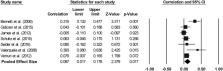
          Population aging has prompted considerable interest in identifying modifiable factors that may help protect the brain and its functions. Collectively, epidemiological studies show that leisure activities with high mental and social demands are linked with better cognition in old age. The extent to which socio-intellectual activities relate to the brain’s structure is, however, not yet fully understood. This systematic review and meta-analysis summarizes magnetic resonance imaging studies that have investigated whether cognitive and social activities correlate with measures of gray and white matter volume, white matter microstructure and white matter lesions. Across eighteen included studies (total n = 8429), activity levels were associated with whole-brain white matter volume, white matter lesions and regional gray matter volume, although effect sizes were small. No associations were found for global gray matter volume and the evidence concerning white matter microstructure was inconclusive. While the causality of the reviewed associations needs to be established, our findings implicate socio-intellectual activity levels as promising targets for interventions aimed at promoting healthy brain aging.

          Related collections

          Most cited references73

          • Record: found
          • Abstract: found
          • Article: not found

          Brain-derived neurotrophic factor is associated with age-related decline in hippocampal volume.

          Hippocampal volume shrinks in late adulthood, but the neuromolecular factors that trigger hippocampal decay in aging humans remains a matter of speculation. In rodents, brain-derived neurotrophic factor (BDNF) promotes the growth and proliferation of cells in the hippocampus and is important in long-term potentiation and memory formation. In humans, circulating levels of BDNF decline with advancing age, and a genetic polymorphism for BDNF has been related to gray matter volume loss in old age. In this study, we tested whether age-related reductions in serum levels of BDNF would be related to shrinkage of the hippocampus and memory deficits in older adults. Hippocampal volume was acquired by automated segmentation of magnetic resonance images in 142 older adults without dementia. The caudate nucleus was also segmented and examined in relation to levels of serum BDNF. Spatial memory was tested using a paradigm in which memory load was parametrically increased. We found that increasing age was associated with smaller hippocampal volumes, reduced levels of serum BDNF, and poorer memory performance. Lower levels of BDNF were associated with smaller hippocampi and poorer memory, even when controlling for the variation related to age. In an exploratory mediation analysis, hippocampal volume mediated the age-related decline in spatial memory and BDNF mediated the age-related decline in hippocampal volume. Caudate nucleus volume was unrelated to BDNF levels or spatial memory performance. Our results identify serum BDNF as a significant factor related to hippocampal shrinkage and memory decline in late adulthood.
            Bookmark
            • Record: found
            • Abstract: found
            • Article: not found

            Age-related changes in grey and white matter structure throughout adulthood

            Normal ageing is associated with gradual brain atrophy. Determining spatial and temporal patterns of change can help shed light on underlying mechanisms. Neuroimaging provides various measures of brain structure that can be used to assess such age-related change but studies to date have typically considered single imaging measures. Although there is consensus on the notion that brain structure deteriorates with age, evidence on the precise time course and spatial distribution of changes is mixed. We assessed grey matter (GM) and white matter (WM) structure in a group of 66 adults aged between 23 and 81. Multimodal imaging measures included voxel-based morphometry (VBM)-style analysis of GM and WM volume and diffusion tensor imaging (DTI) metrics of WM microstructure. We found widespread reductions in GM volume from middle age onwards but earlier reductions in GM were detected in frontal cortex. Widespread age-related deterioration in WM microstructure was detected from young adulthood onwards. WM decline was detected earlier and more sensitively using DTI-based measures of microstructure than using markers of WM volume derived from conventional T1-weighted imaging.
              Bookmark
              • Record: found
              • Abstract: found
              • Article: not found

              Longitudinal pattern of regional brain volume change differentiates normal aging from MCI.

              Neuroimaging measures have potential as surrogate markers of disease through identification of consistent features that occur prior to clinical symptoms. Despite numerous investigations, especially in relation to the transition to clinical impairment, the regional pattern of brain changes in clinically normal older adults has not been established. We predict that the regions that show early pathologic changes in association with Alzheimer disease will show accelerated volume loss in mild cognitive impairment (MCI) compared to normal aging. Through the Baltimore Longitudinal Study of Aging, we prospectively evaluated 138 nondemented individuals (age 64-86 years) annually for up to 10 consecutive years. Eighteen participants were diagnosed with MCI over the course of the study. Mixed-effects regression was used to compare regional brain volume trajectories of clinically normal individuals to those with MCI based on a total of 1,017 observations. All investigated volumes declined with normal aging (p < 0.05). Accelerated change with age was observed for ventricular CSF (vCSF), frontal gray matter, superior, middle, and medial frontal, and superior parietal regions (p < or = 0.04). The MCI group showed accelerated changes compared to normal controls in whole brain volume, vCSF, temporal gray matter, and orbitofrontal and temporal association cortices, including the hippocampus (p < or = 0.04). Although age-related regional volume loss is apparent and widespread in nondemented individuals, mild cognitive impairment is associated with a unique pattern of structural vulnerability reflected in differential volume loss in specific regions. Early identification of patterns of abnormality is of fundamental importance for detecting disease onset and tracking progression.
                Bookmark

                Author and article information

                Contributors
                Journal
                Neurosci Biobehav Rev
                Neurosci Biobehav Rev
                Neuroscience and Biobehavioral Reviews
                Pergamon Press
                0149-7634
                1873-7528
                1 October 2018
                October 2018
                : 93
                : 71-84
                Affiliations
                [a ]Department of Psychiatry, University of Oxford, Warneford Hospital, Warneford Lane, Oxford, OX3 7JX, United Kingdom
                [b ]Wellcome Centre for Integrative Neuroimaging, Oxford Centre for Human Brain Activity, Department of Psychaitry, University of Oxford, Warneford Hospital, Warneford Lane, Oxford, OX3 7JX, United Kingdom
                [c ]Global Brain Health Institute, Memory and Aging Center, Department of Neurology, University of California, San Francisco, CA, 94158, USA
                Author notes
                [* ]Corresponding author at: Wellcome Centre for Integrative Neuroimaging, Oxford Centre for Human Brain Activity, Department of Psychaitry, University of Oxford, Warneford Hospital, Warneford Lane, Oxford, OX3 7JX, United Kingdom. claire.sexton@ 123456psych.ox.ac.uk
                Article
                S0149-7634(18)30245-8
                10.1016/j.neubiorev.2018.06.012
                6562200
                29940239
                0bff13c5-f1d0-45fa-8c16-00c20beb45c5
                © 2018 The Authors

                This is an open access article under the CC BY license (http://creativecommons.org/licenses/by/4.0/).

                History
                : 3 April 2018
                : 13 June 2018
                : 15 June 2018
                Categories
                Article

                Neurosciences
                brain,social activity,cognitive activity,aging,magnetic resonance imaging,voxel-based morphometry,region-of-interest,gray matter,white matter

                Comments

                Comment on this article