22
views
0
recommends
+1 Recommend
0 collections
    0
    shares
      • Record: found
      • Abstract: found
      • Article: found
      Is Open Access

      Insect Responses to Linearly Polarized Reflections: Orphan Behaviors Without Neural Circuits

      review-article

      Read this article at

      Bookmark
          There is no author summary for this article yet. Authors can add summaries to their articles on ScienceOpen to make them more accessible to a non-specialist audience.

          Abstract

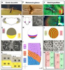
          The e-vector orientation of linearly polarized light represents an important visual stimulus for many insects. Especially the detection of polarized skylight by many navigating insect species is known to improve their orientation skills. While great progress has been made towards describing both the anatomy and function of neural circuit elements mediating behaviors related to navigation, relatively little is known about how insects perceive non-celestial polarized light stimuli, like reflections off water, leaves, or shiny body surfaces. Work on different species suggests that these behaviors are not mediated by the “Dorsal Rim Area” (DRA), a specialized region in the dorsal periphery of the adult compound eye, where ommatidia contain highly polarization-sensitive photoreceptor cells whose receptive fields point towards the sky. So far, only few cases of polarization-sensitive photoreceptors have been described in the ventral periphery of the insect retina. Furthermore, both the structure and function of those neural circuits connecting to these photoreceptor inputs remain largely uncharacterized. Here we review the known data on non-celestial polarization vision from different insect species (dragonflies, butterflies, beetles, bugs and flies) and present three well-characterized examples for functionally specialized non-DRA detectors from different insects that seem perfectly suited for mediating such behaviors. Finally, using recent advances from circuit dissection in Drosophila melanogaster, we discuss what types of potential candidate neurons could be involved in forming the underlying neural circuitry mediating non-celestial polarization vision.

          Related collections

          Most cited references100

          • Record: found
          • Abstract: found
          • Article: not found

          Structural origin of circularly polarized iridescence in jeweled beetles.

          The iridescent metallic green beetle, Chrysina gloriosa, which selectively reflects left circularly polarized light, possesses an exoskeleton decorated by hexagonal cells (approximately 10 microm) that coexist with pentagons and heptagons. The fraction of hexagons decreases with an increase in curvature. In bright field microscopy, each cell contains a bright yellow core, placed in a greenish cell with yellowish border, but the core disappears in dark field. With use of confocal microscopy, we observe that these cells consist of nearly concentric nested arcs that lie on the surface of a shallow cone. We infer that the patterns are structurally and optically analogous to the focal conic domains formed spontaneously on the free surface of a cholesteric liquid crystal. These textures provide the basis for the morphogenesis as well as key insights for emulating the intricate optical response of the exoskeleton of scarab beetles.
            Bookmark
            • Record: found
            • Abstract: found
            • Article: not found

            Stochastic spineless expression creates the retinal mosaic for colour vision.

            Drosophila colour vision is achieved by R7 and R8 photoreceptor cells present in every ommatidium. The fly retina contains two types of ommatidia, called 'pale' and 'yellow', defined by different rhodopsin pairs expressed in R7 and R8 cells. Similar to the human cone photoreceptors, these ommatidial subtypes are distributed stochastically in the retina. The choice between pale versus yellow ommatidia is made in R7 cells, which then impose their fate onto R8. Here we report that the Drosophila dioxin receptor Spineless is both necessary and sufficient for the formation of the ommatidial mosaic. A short burst of spineless expression at mid-pupation in a large subset of R7 cells precedes rhodopsin expression. In spineless mutants, all R7 and most R8 cells adopt the pale fate, whereas overexpression of spineless is sufficient to induce the yellow R7 fate. Therefore, this study suggests that the entire retinal mosaic required for colour vision is defined by the stochastic expression of a single transcription factor, Spineless.
              Bookmark
              • Record: found
              • Abstract: found
              • Article: not found

              Maplike representation of celestial E-vector orientations in the brain of an insect.

              For many insects, the polarization pattern of the blue sky serves as a compass cue for spatial navigation. E-vector orientations are detected by photoreceptors in a dorsal rim area of the eye. Polarized-light signals from both eyes are finally integrated in the central complex, a brain area consisting of two subunits, the protocerebral bridge and the central body. Here we show that a topographic representation of zenithal E-vector orientations underlies the columnar organization of the protocerebral bridge in a locust. The maplike arrangement is highly suited to signal head orientation under the open sky.
                Bookmark

                Author and article information

                Contributors
                Journal
                Front Cell Neurosci
                Front Cell Neurosci
                Front. Cell. Neurosci.
                Frontiers in Cellular Neuroscience
                Frontiers Media S.A.
                1662-5102
                20 March 2018
                2018
                : 12
                : 50
                Affiliations
                [1]Division of Neurobiology, Institut für Biology, Fachbereich Biologie, Chemie & Pharmazie, Freie Universität Berlin , Berlin, Germany
                Author notes

                Edited by: Vilaiwan M. Fernandes, New York University, United States

                Reviewed by: Gregor Belusic, University of Ljubljana, Slovenia; Abderrahman Khila, UMR5203 Institut de Génomique Fonctionnelle (IGF), France

                *Correspondence: Mathias F. Wernet mathias.wernet@ 123456fu-berlin.de
                Article
                10.3389/fncel.2018.00050
                5870057
                29615868
                0c23962a-f191-4c2f-8780-2bda74cccc2c
                Copyright © 2018 Heinloth, Uhlhorn and Wernet.

                This is an open-access article distributed under the terms of the Creative Commons Attribution License (CC BY). The use, distribution or reproduction in other forums is permitted, provided the original author(s) and the copyright owner are credited and that the original publication in this journal is cited, in accordance with accepted academic practice. No use, distribution or reproduction is permitted which does not comply with these terms.

                History
                : 19 November 2017
                : 15 February 2018
                Page count
                Figures: 4, Tables: 0, Equations: 0, References: 106, Pages: 13, Words: 11123
                Funding
                Funded by: Deutsche Forschungsgemeinschaft 10.13039/501100001659
                Award ID: WE 5761/2-1, SFB958
                Categories
                Neuroscience
                Review

                Neurosciences
                insect vision,polarized light,behavior,orientation,water detection,neuroethology,visual ecology,neural circuits

                Comments

                Comment on this article