43
views
0
recommends
+1 Recommend
0 collections
    0
    shares
      • Record: found
      • Abstract: found
      • Article: not found

      Polyglutamine-expanded androgen receptor interferes with TFEB to elicit pathological autophagy defects in SBMA

      research-article

      Read this article at

      ScienceOpenPublisherPMC
      Bookmark
          There is no author summary for this article yet. Authors can add summaries to their articles on ScienceOpen to make them more accessible to a non-specialist audience.

          Abstract

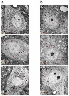
          Macroautophagy (hereafter autophagy) is a key pathway in neurodegeneration. Despite protective actions, autophagy may contribute to neuron demise, when dysregulated. Here we considered X-linked spinal and bulbar muscular atrophy (SBMA), a repeat disorder caused by polyglutamine-expanded androgen receptor (polyQ-AR). We found that polyQ-AR reduced long-term protein turnover and impaired autophagic flux in motor neuron-like cells. Ultrastructural analysis of SBMA mice revealed a block in autophagy pathway progression. We considered the transcriptional regulation of autophagy, and observed a functionally significant physical interaction between transcription factor EB (TFEB) and AR. Normal AR promoted, but polyQ-AR interfered with TFEB transactivation. To evaluate physiological relevance, we reprogrammed patient fibroblasts to induced pluripotent stem cells, and then to neuronal precursor cells (NPCs). We compared multiple SBMA NPC lines, and documented metabolic and autophagic flux defects that could be rescued by TFEB. Our results indicate that polyQ-AR diminishes TFEB function to impair autophagy and promote SBMA pathogenesis.

          Related collections

          Most cited references28

          • Record: found
          • Abstract: found
          • Article: not found

          Guidelines for the use and interpretation of assays for monitoring autophagy.

          In 2008 we published the first set of guidelines for standardizing research in autophagy. Since then, research on this topic has continued to accelerate, and many new scientists have entered the field. Our knowledge base and relevant new technologies have also been expanding. Accordingly, it is important to update these guidelines for monitoring autophagy in different organisms. Various reviews have described the range of assays that have been used for this purpose. Nevertheless, there continues to be confusion regarding acceptable methods to measure autophagy, especially in multicellular eukaryotes. A key point that needs to be emphasized is that there is a difference between measurements that monitor the numbers or volume of autophagic elements (e.g., autophagosomes or autolysosomes) at any stage of the autophagic process vs. those that measure flux through the autophagy pathway (i.e., the complete process); thus, a block in macroautophagy that results in autophagosome accumulation needs to be differentiated from stimuli that result in increased autophagic activity, defined as increased autophagy induction coupled with increased delivery to, and degradation within, lysosomes (in most higher eukaryotes and some protists such as Dictyostelium) or the vacuole (in plants and fungi). In other words, it is especially important that investigators new to the field understand that the appearance of more autophagosomes does not necessarily equate with more autophagy. In fact, in many cases, autophagosomes accumulate because of a block in trafficking to lysosomes without a concomitant change in autophagosome biogenesis, whereas an increase in autolysosomes may reflect a reduction in degradative activity. Here, we present a set of guidelines for the selection and interpretation of methods for use by investigators who aim to examine macroautophagy and related processes, as well as for reviewers who need to provide realistic and reasonable critiques of papers that are focused on these processes. These guidelines are not meant to be a formulaic set of rules, because the appropriate assays depend in part on the question being asked and the system being used. In addition, we emphasize that no individual assay is guaranteed to be the most appropriate one in every situation, and we strongly recommend the use of multiple assays to monitor autophagy. In these guidelines, we consider these various methods of assessing autophagy and what information can, or cannot, be obtained from them. Finally, by discussing the merits and limits of particular autophagy assays, we hope to encourage technical innovation in the field.
            Bookmark
            • Record: found
            • Abstract: found
            • Article: not found

            A model for neural development and treatment of Rett syndrome using human induced pluripotent stem cells.

            Autism spectrum disorders (ASD) are complex neurodevelopmental diseases in which different combinations of genetic mutations may contribute to the phenotype. Using Rett syndrome (RTT) as an ASD genetic model, we developed a culture system using induced pluripotent stem cells (iPSCs) from RTT patients' fibroblasts. RTT patients' iPSCs are able to undergo X-inactivation and generate functional neurons. Neurons derived from RTT-iPSCs had fewer synapses, reduced spine density, smaller soma size, altered calcium signaling and electrophysiological defects when compared to controls. Our data uncovered early alterations in developing human RTT neurons. Finally, we used RTT neurons to test the effects of drugs in rescuing synaptic defects. Our data provide evidence of an unexplored developmental window, before disease onset, in RTT syndrome where potential therapies could be successfully employed. Our model recapitulates early stages of a human neurodevelopmental disease and represents a promising cellular tool for drug screening, diagnosis and personalized treatment. Copyright © 2010 Elsevier Inc. All rights reserved.
              Bookmark
              • Record: found
              • Abstract: found
              • Article: not found

              TFEB-mediated autophagy rescues midbrain dopamine neurons from α-synuclein toxicity.

              The aggregation of α-synuclein plays a major role in Parkinson disease (PD) pathogenesis. Recent evidence suggests that defects in the autophagy-mediated clearance of α-synuclein contribute to the progressive loss of nigral dopamine neurons. Using an in vivo model of α-synuclein toxicity, we show that the PD-like neurodegenerative changes induced by excess cellular levels of α-synuclein in nigral dopamine neurons are closely linked to a progressive decline in markers of lysosome function, accompanied by cytoplasmic retention of transcription factor EB (TFEB), a major transcriptional regulator of the autophagy-lysosome pathway. The changes in lysosomal function, observed in the rat model as well as in human PD midbrain, were reversed by overexpression of TFEB, which afforded robust neuroprotection via the clearance of α-synuclein oligomers, and were aggravated by microRNA-128-mediated repression of TFEB in both A9 and A10 dopamine neurons. Delayed activation of TFEB function through inhibition of mammalian target of rapamycin blocked α-synuclein induced neurodegeneration and further disease progression. The results provide a mechanistic link between α-synuclein toxicity and impaired TFEB function, and highlight TFEB as a key player in the induction of α-synuclein-induced toxicity and PD pathogenesis, thus identifying TFEB as a promising target for therapies aimed at neuroprotection and disease modification in PD.
                Bookmark

                Author and article information

                Journal
                9809671
                21092
                Nat Neurosci
                Nat. Neurosci.
                Nature neuroscience
                1097-6256
                1546-1726
                24 September 2014
                10 August 2014
                September 2014
                01 March 2015
                : 17
                : 9
                : 1180-1189
                Affiliations
                [1 ]Department of Pediatrics, University of California, San Diego; La Jolla, CA 92093, USA
                [2 ]Department of Cellular & Molecular Medicine, University of California, San Diego; La Jolla, CA 92093, USA
                [3 ]Center on Human Development & Disability, University of Washington, Seattle, WA 98195, USA
                [4 ]Department of Neurology, University of Washington, Seattle, WA 98195, USA
                [5 ]Department of Neurosciences, University of California, San Diego; La Jolla, CA 92093, USA
                [6 ]Division of Biological Sciences, University of California, San Diego; La Jolla, CA 92093, USA
                [7 ]Institute for Genomic Medicine, University of California, San Diego; La Jolla, CA 92093, USA
                [8 ]Sanford Consortium for Regenerative Medicine, University of California, San Diego; La Jolla, CA 92093, USA
                [9 ]Rady Children's Hospital, San Diego, CA 92123, USA
                Author notes
                Corresponding author: Albert La Spada, MD, PhD, Cellular & Molecular Medicine, Neurosciences, and Pediatrics, University of California, San Diego, 9500 Gilman Drive, MC 0642, La Jolla, CA 92093-0642, (858)-246-0148 [ph.], alaspada@ 123456ucsd.edu
                [*]

                These authors contributed equally to this work.

                Article
                NIHMS614692
                10.1038/nn.3787
                4180729
                25108912
                0c381a9a-cc43-4c9d-a4d3-f69bf905ffb9
                History
                Categories
                Article

                Neurosciences
                Neurosciences

                Comments

                Comment on this article