47
views
0
recommends
+1 Recommend
0 collections
    0
    shares
      • Record: found
      • Abstract: found
      • Article: found
      Is Open Access

      HIV-1 Vpr activates the G 2 checkpoint through manipulation of the ubiquitin proteasome system

      research-article

      Read this article at

      Bookmark
          There is no author summary for this article yet. Authors can add summaries to their articles on ScienceOpen to make them more accessible to a non-specialist audience.

          Abstract

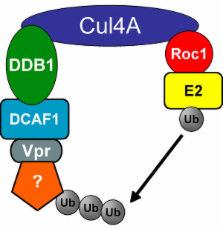
          HIV-1 Vpr is a viral accessory protein that activates ATR through the induction of DNA replication stress. ATR activation results in cell cycle arrest in G 2 and induction of apoptosis. In the present study, we investigate the role of the ubiquitin/proteasome system (UPS) in the above activity of Vpr. We report that the general function of the UPS is required for Vpr to induce G 2 checkpoint activation, as incubation of Vpr-expressing cells with proteasome inhibitors abolishes this effect. We further investigated in detail the specific E3 ubiquitin ligase subunits that Vpr manipulates. We found that Vpr binds to the DCAF1 subunit of a cullin 4a/DDB1 E3 ubiquitin ligase. The carboxy-terminal domain Vpr(R80A) mutant, which is able to bind DCAF1, is inactive in checkpoint activation and has dominant-negative character. In contrast, the mutation Q65R, in the leucine-rich domain of Vpr that mediates DCAF1 binding, results in an inactive Vpr devoid of dominant negative behavior. Thus, the interaction of Vpr with DCAF1 is required, but not sufficient, for Vpr to cause G 2 arrest. We propose that Vpr recruits, through its carboxy terminal domain, an unknown cellular factor that is required for G 2-to-M transition. Recruitment of this factor leads to its ubiquitination and degradation, resulting in failure to enter mitosis.

          Related collections

          Most cited references31

          • Record: found
          • Abstract: found
          • Article: not found

          Molecular architecture and assembly of the DDB1-CUL4A ubiquitin ligase machinery.

          Protein ubiquitination is a common form of post-translational modification that regulates a broad spectrum of protein substrates in diverse cellular pathways. Through a three-enzyme (E1-E2-E3) cascade, the attachment of ubiquitin to proteins is catalysed by the E3 ubiquitin ligase, which is best represented by the superfamily of the cullin-RING complexes. Conserved from yeast to human, the DDB1-CUL4-ROC1 complex is a recently identified cullin-RING ubiquitin ligase, which regulates DNA repair, DNA replication and transcription, and can also be subverted by pathogenic viruses to benefit viral infection. Lacking a canonical SKP1-like cullin adaptor and a defined substrate recruitment module, how the DDB1-CUL4-ROC1 E3 apparatus is assembled for ubiquitinating various substrates remains unclear. Here we present crystallographic analyses of the virally hijacked form of the human DDB1-CUL4A-ROC1 machinery, which show that DDB1 uses one beta-propeller domain for cullin scaffold binding and a variably attached separate double-beta-propeller fold for substrate presentation. Through tandem-affinity purification of human DDB1 and CUL4A complexes followed by mass spectrometry analysis, we then identify a novel family of WD40-repeat proteins, which directly bind to the double-propeller fold of DDB1 and serve as the substrate-recruiting module of the E3. Together, our structural and proteomic results reveal the structural mechanisms and molecular logic underlying the assembly and versatility of a new family of cullin-RING E3 complexes.
            Bookmark
            • Record: found
            • Abstract: found
            • Article: not found

            ATR regulates fragile site stability.

            Conditions that partially inhibit DNA replication induce expression of common fragile sites. These sites form gaps and breaks on metaphase chromosomes and are deleted and rearranged in many tumors. Yet, the mechanism of fragile site expression has been elusive. We demonstrate that the replication checkpoint kinase ATR, but not ATM, is critical for maintenance of fragile site stability. ATR deficiency results in fragile site expression with and without addition of replication inhibitors. Thus, we propose that fragile sites are unreplicated chromosomal regions resulting from stalled forks that escape the ATR replication checkpoint. These findings have important implications for understanding both the mechanism of fragile site instability and the consequences of stalled replication in mammalian cells.
              Bookmark
              • Record: found
              • Abstract: found
              • Article: not found

              A Novel Human WD Protein, h-βTrCP, that Interacts with HIV-1 Vpu Connects CD4 to the ER Degradation Pathway through an F-Box Motif

              HIV-1 Vpu interacts with CD4 in the endoplasmic reticulum and triggers CD4 degradation, presumably by proteasomes. Human beta TrCP identified by interaction with Vpu connects CD4 to this proteolytic machinery, and CD4-Vpu-beta TrCP ternary complexes have been detected by coimmunoprecipitation. beta TrCP binding to Vpu and its recruitment to membranes require two phosphoserine residues in Vpu essential for CD4 degradation. In beta TrCP, WD repeats at the C terminus mediate binding to Vpu, and an F box near the N terminus is involved in interaction with Skp1p, a targeting factor for ubiquitin-mediated proteolysis. An F-box deletion mutant of beta TrCP had a dominant-negative effect on Vpu-mediated CD4 degradation. These data suggest that beta TrCP and Skp1p represent components of a novel ER-associated protein degradation pathway that mediates CD4 proteolysis.
                Bookmark

                Author and article information

                Journal
                Virol J
                Virology Journal
                BioMed Central (London )
                1743-422X
                2007
                8 June 2007
                : 4
                : 57
                Affiliations
                [1 ]Division of Cell Biology and Immunology, Department of Pathology, University of Utah School of Medicine, 15 North Medical Drive East #2100 – Room 2520, Salt Lake City, UT 84112, USA
                [2 ]Laboratório de Farmacologia Molecular (CP 04536), Faculdade de Saude, Universidade de Brasília, 70919-970 Brasília, DF, Brazil
                Article
                1743-422X-4-57
                10.1186/1743-422X-4-57
                1904188
                17559673
                0cf0863c-71b0-4906-a820-beca63cb12cc
                Copyright © 2007 DeHart et al; licensee BioMed Central Ltd.

                This is an Open Access article distributed under the terms of the Creative Commons Attribution License ( http://creativecommons.org/licenses/by/2.0), which permits unrestricted use, distribution, and reproduction in any medium, provided the original work is properly cited.

                History
                : 4 June 2007
                : 8 June 2007
                Categories
                Research

                Microbiology & Virology
                Microbiology & Virology

                Comments

                Comment on this article