13
views
0
recommends
+1 Recommend
0 collections
    0
    shares
      • Record: found
      • Abstract: found
      • Article: found
      Is Open Access

      Interest of Ultrasonographic Assessment of Diaphragmatic Function in Cardiac Rehabilitation Center: A Case Report

      case-report
      , MD, PhD, , PhD, , MD
      Medicine
      Wolters Kluwer Health

      Read this article at

      Bookmark
          There is no author summary for this article yet. Authors can add summaries to their articles on ScienceOpen to make them more accessible to a non-specialist audience.

          Abstract

          Diaphragmatic paresis is a rare but recognized complication of atrial fibrillation ablation.

          A 59-year-old woman experiencing dyspnea in supine position and for minimal effort was admitted in a cardiac rehabilitation center. One month before, she was referred to a cardiac center to ablation of paroxysmal atrial fibrillation. After the procedure, the patient developed respiratory failure attributed to aspiration pneumonia and requiring mechanical ventilation.

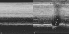
          At admission in the rehabilitation center, M-mode ultrasonography reported an absence of movement of the right hemidiaphragm during quiet breathing and a paradoxical movement during voluntary sniffing.

          Chest ultrasonography can be useful to detect diaphragmatic dysfunction in patients suffering from dyspnea, at admission in a cardiac rehabilitation center. Its use should be envisaged more frequently.

          Related collections

          Most cited references9

          • Record: found
          • Abstract: found
          • Article: not found

          Diaphragmatic motion studied by m-mode ultrasonography: methods, reproducibility, and normal values.

          Although diaphragmatic motion is readily studied by ultrasonography, the procedure remains poorly codified. The aim of this prospective study was to determine the reference values for diaphragmatic motion as recorded by M-mode ultrasonography. Two hundred ten healthy adult subjects (150 men, 60 women) were investigated. Both sides of the posterior diaphragm were identified, and M-mode was used to display the movement of the anatomical structures. Examinations were performed during quiet breathing, voluntary sniffing, and deep breathing. Diaphragmatic excursions were measured from the M-mode sonographic images. In addition, the reproducibility (inter- and intra-observer) was assessed. Right and left diaphragmatic motions were successfully assessed during quiet breathing in all subjects. During voluntary sniffing, the measurement was always possible on the right side, and in 208 of 210 volunteers, on the left side. During deep breathing, an obscuration of the diaphragm by the descending lung was noted in subjects with marked diaphragmatic excursion. Consequently, right diaphragmatic excursion could be measured in 195 of 210 subjects, and left diaphragmatic excursion in only 45 subjects. Finally, normal values of both diaphragmatic excursions were determined. Since the excursions were larger in men than in women, the gender should be taken into account. The lower limit values were close to 0.9 cm for women and 1 cm for men during quiet breathing, 1.6 cm for women and 1.8 cm for men during voluntary sniffing, and 3.7 cm for women and 4.7 cm for men during deep breathing. We demonstrated that M-mode ultrasonography is a reproducible method for assessing hemidiaphragmatic movement.
            Bookmark
            • Record: found
            • Abstract: found
            • Article: not found

            Diaphragmatic paralysis: the use of M mode ultrasound for diagnosis in adults.

            Retrospective, case series.
              Bookmark
              • Record: found
              • Abstract: found
              • Article: not found

              Inspiratory muscle training for diaphragm dysfunction after cardiac surgery.

              Diaphragm dysfunction is a complication of cardiac surgery with partial or absent spontaneous recovery in most cases. Surgical diaphragm plication represents the only option when symptoms persist. Because training improves functional nerve recovery after a nerve lesion, we hypothesized that early diaphragm muscle training may be beneficial.
                Bookmark

                Author and article information

                Journal
                Medicine (Baltimore)
                Medicine (Baltimore)
                MEDI
                Medicine
                Wolters Kluwer Health
                0025-7974
                1536-5964
                May 2015
                21 May 2015
                : 94
                : 19
                : e801
                Affiliations
                From the UMRMD2, Aix-Marseille University and IRBA, Marseille (AB, GC); Unité de Réhabilitation Cardiologique, Hôpital Léon-Bérard, Hyères, France (LP).
                Author notes
                Correspondence: Alain Boussuges, UMRMD2, Faculté de Médecine Nord, Marseille, France (e-mail: alain.boussuges@ 123456univ-amu.fr ).
                Article
                00801
                10.1097/MD.0000000000000801
                4602564
                25984664
                0d2100e2-c5bd-4e0b-a382-75dcc4e3311f
                Copyright © 2015 Wolters Kluwer Health, Inc. All rights reserved.

                This is an open access article distributed under the Creative Commons Attribution License 4.0, which permits unrestricted use, distribution, and reproduction in any medium, provided the original work is properly cited. http://creativecommons.org/licenses/by/4.0

                History
                : 20 February 2015
                : 31 March 2015
                : 24 March 2015
                Categories
                6300
                Article
                Clinical Case Report
                Custom metadata
                TRUE

                Comments

                Comment on this article