8
views
0
recommends
+1 Recommend
0 collections
    0
    shares
      • Record: found
      • Abstract: found
      • Article: found
      Is Open Access

      Structural basis for electron transport mechanism of complex I-like photosynthetic NAD(P)H dehydrogenase

      research-article

      Read this article at

      Bookmark
          There is no author summary for this article yet. Authors can add summaries to their articles on ScienceOpen to make them more accessible to a non-specialist audience.

          Abstract

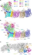
          NAD(P)H dehydrogenase-like (NDH) complex NDH-1L of cyanobacteria plays a crucial role in cyclic electron flow (CEF) around photosystem I and respiration processes. NDH-1L couples the electron transport from ferredoxin (Fd) to plastoquinone (PQ) and proton pumping from cytoplasm to the lumen that drives the ATP production. NDH-1L-dependent CEF increases the ATP/NADPH ratio, and is therefore pivotal for oxygenic phototrophs to function under stress. Here we report two structures of NDH-1L from Thermosynechococcus elongatus BP-1, in complex with one Fd and an endogenous PQ, respectively. Our structures represent the complete model of cyanobacterial NDH-1L, revealing the binding manner of NDH-1L with Fd and PQ, as well as the structural elements crucial for proper functioning of the NDH-1L complex. Together, our data provides deep insights into the electron transport from Fd to PQ, and its coupling with proton translocation in NDH-1L.

          Abstract

          NAD(P)H dehydrogenase-like complex NDH-1L couples the electron transport from ferredoxin (Fd) to plastoquinone (PQ) and proton pumping from cytoplasm to the lumen. Here authors report two structures of NDH-1L from Thermosynechococcus elongatus BP-1, in complex with one Fd and an endogenous PQ, respectively.

          Related collections

          Most cited references51

          • Record: found
          • Abstract: found
          • Article: not found

          Natural engineering principles of electron tunnelling in biological oxidation-reduction.

          We have surveyed proteins with known atomic structure whose function involves electron transfer; in these, electrons can travel up to 14 A between redox centres through the protein medium. Transfer over longer distances always involves a chain of cofactors. This redox centre proximity alone is sufficient to allow tunnelling of electrons at rates far faster than the substrate redox reactions it supports. Consequently, there has been no necessity for proteins to evolve optimized routes between redox centres. Instead, simple geometry enables rapid tunnelling to high-energy intermediate states. This greatly simplifies any analysis of redox protein mechanisms and challenges the need to postulate mechanisms of superexchange through redox centres or the maintenance of charge neutrality when investigating electron-transfer reactions. Such tunnelling also allows sequential electron transfer in catalytic sites to surmount radical transition states without involving the movement of hydride ions, as is generally assumed. The 14 A or less spacing of redox centres provides highly robust engineering for electron transfer, and may reflect selection against designs that have proved more vulnerable to mutations during the course of evolution.
            Bookmark
            • Record: found
            • Abstract: found
            • Article: not found

            Crystal structure of the entire respiratory complex I

            Complex I is the first and largest enzyme of the respiratory chain, playing a central role in cellular energy production by coupling electron transfer between NADH and ubiquinone to proton translocation. It is implicated in many common human neurodegenerative diseases. Here we report the first crystal structure of the entire, intact complex I (from T. thermophilus) at 3.3 Å resolution. The structure of the 536 kDa complex comprises 16 different subunits with 64 transmembrane helices and 9 Fe-S clusters. The core fold of subunit Nqo8 (NuoH/ND1) is, unexpectedly, similar to a half-channel of the antiporter-like subunits. Small subunits nearby form a linked second half-channel, thus completing the fourth proton translocation pathway, in addition to the channels in three antiporter-like subunits. The quinone-binding site is unusually long, narrow and enclosed. The quinone headgroup binds at the deep end of this chamber, near cluster N2. Strikingly, the chamber is linked to the fourth channel by a “funnel” of charged residues. The link continues over the entire membrane domain as a remarkable flexible central axis of charged and polar residues. It likely plays a leading role in the propagation of conformational changes, aided by coupling elements. The structure suggests that a unique, out-of-the-membrane quinone reaction chamber allows the redox energy to drive concerted long-range conformational changes in the four antiporter-like domains, resulting in translocation of four protons per cycle.
              Bookmark
              • Record: found
              • Abstract: found
              • Article: not found

              Cyclic electron flow around photosystem I is essential for photosynthesis.

              Photosynthesis provides at least two routes through which light energy can be used to generate a proton gradient across the thylakoid membrane of chloroplasts, which is subsequently used to synthesize ATP. In the first route, electrons released from water in photosystem II (PSII) are eventually transferred to NADP+ by way of photosystem I (PSI). This linear electron flow is driven by two photochemical reactions that function in series. The cytochrome b6f complex mediates electron transport between the two photosystems and generates the proton gradient (DeltapH). In the second route, driven solely by PSI, electrons can be recycled from either reduced ferredoxin or NADPH to plastoquinone, and subsequently to the cytochrome b6f complex. Such cyclic flow generates DeltapH and thus ATP without the accumulation of reduced species. Whereas linear flow from water to NADP+ is commonly used to explain the function of the light-dependent reactions of photosynthesis, the role of cyclic flow is less clear. In higher plants cyclic flow consists of two partially redundant pathways. Here we have constructed mutants in Arabidopsis thaliana in which both PSI cyclic pathways are impaired, and present evidence that cyclic flow is essential for efficient photosynthesis.
                Bookmark

                Author and article information

                Contributors
                hlmi@sibs.ac.cn
                xzzhang@ibp.ac.cn
                meili@ibp.ac.cn
                Journal
                Nat Commun
                Nat Commun
                Nature Communications
                Nature Publishing Group UK (London )
                2041-1723
                30 January 2020
                30 January 2020
                2020
                : 11
                : 610
                Affiliations
                [1 ]ISNI 0000 0004 1792 5640, GRID grid.418856.6, National Laboratory of Biomacromolecules, CAS Center for Excellence in Biomacromolecules, Institute of Biophysics, Chinese Academy of Sciences, ; Beijing, 100101 PR China
                [2 ]ISNI 0000 0004 1797 8419, GRID grid.410726.6, University of Chinese Academy of Sciences, ; Beijing, 100049 PR China
                [3 ]ISNI 0000 0004 0467 2285, GRID grid.419092.7, National Key Laboratory of Plant Molecular Genetics, Institute of Plant Physiology and Ecology, Shanghai Institutes for Biological Sciences, Chinese Academy of Science, ; Shanghai, 200032 PR China
                [4 ]ISNI 0000 0004 1792 5640, GRID grid.418856.6, Center for Biological Imaging, CAS Center for Excellence in Biomacromolecules, Institute of Biophysics, Chinese Academy of Sciences, ; Beijing, 100101 PR China
                Author information
                http://orcid.org/0000-0002-9672-4649
                http://orcid.org/0000-0002-1229-3950
                http://orcid.org/0000-0002-0114-0270
                http://orcid.org/0000-0002-1742-2360
                Article
                14456
                10.1038/s41467-020-14456-0
                6992706
                32001694
                0d97b2ba-6e52-4555-a7ea-ca87ae84fadf
                © The Author(s) 2020

                Open Access This article is licensed under a Creative Commons Attribution 4.0 International License, which permits use, sharing, adaptation, distribution and reproduction in any medium or format, as long as you give appropriate credit to the original author(s) and the source, provide a link to the Creative Commons license, and indicate if changes were made. The images or other third party material in this article are included in the article’s Creative Commons license, unless indicated otherwise in a credit line to the material. If material is not included in the article’s Creative Commons license and your intended use is not permitted by statutory regulation or exceeds the permitted use, you will need to obtain permission directly from the copyright holder. To view a copy of this license, visit http://creativecommons.org/licenses/by/4.0/.

                History
                : 25 September 2019
                : 9 January 2020
                Funding
                Funded by: FundRef https://doi.org/10.13039/501100002855, Ministry of Science and Technology of the People's Republic of China (Chinese Ministry of Science and Technology);
                Award ID: 2016YFA0502900
                Award ID: 2017YFA0504700
                Award ID: 2017YFA0503702
                Award Recipient :
                Funded by: FundRef https://doi.org/10.13039/501100001809, National Natural Science Foundation of China (National Science Foundation of China);
                Award ID: 31970264, 31600609
                Award ID: 31770257
                Award ID: 31930064, 31770778
                Award Recipient :
                Funded by: Ministry of Science and Technology of the People's Republic of China (Chinese Ministry of Science and Technology)
                Funded by: FundRef https://doi.org/10.13039/501100002367, Chinese Academy of Sciences (CAS);
                Award ID: XDB08030204
                Award ID: XDB27020106, XDB08020302, QYZDB-SSW-SMC005
                Award Recipient :
                Funded by: Ministry of Science and Technology of the People's Republic of China (Chinese Ministry of Science and Technology)
                Categories
                Article
                Custom metadata
                © The Author(s) 2020

                Uncategorized
                membrane proteins,cryoelectron microscopy
                Uncategorized
                membrane proteins, cryoelectron microscopy

                Comments

                Comment on this article