2
views
0
recommends
+1 Recommend
0 collections
    0
    shares
      • Record: found
      • Abstract: found
      • Article: found
      Is Open Access

      Performance of Ultrasound in the Clinical Evaluation of Gout and Hyperuricemia

      research-article

      Read this article at

      Bookmark
          There is no author summary for this article yet. Authors can add summaries to their articles on ScienceOpen to make them more accessible to a non-specialist audience.

          Abstract

          Objective

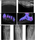
          To evaluate monosodium urate (MSU) crystal deposition and related lesions in the joints of patients with gout and hyperuricemia (HUA) using ultrasound. To explore the association between ultrasound findings and clinical features in gout and HUA.

          Methods

          A total of 202 patients with gout and 43 asymptomatic patients with HUA were included. The clinical data and ultrasonic assessment results were collected and statistically analyzed.

          Results

          Deposition of MSU crystals was found in 25.58% (11/43) of patients with asymptomatic HUA and 76.24% (154/202) of patients with gout. Of the 1,082 joints from patients with gout examined, 33.09% (358/1082) displayed MSU crystal deposition. In the joints with MSU crystal deposition, 77.37% (277/358) had a history of attacks. Among the joints of gouty arthritis, double contour sign (DCS), hyperechoic aggregate (HAG), and tophi were found in 32.65% (159/487), 7.80% (38/487), and 24.64% (120/487) of the joints, respectively. DCS and tophi, but not HAG, increasingly appeared with the extension of gout duration. In patients with more than 15 years of gout history, DCS, Tophi, and HAG were found in 48.18%, 40.00%, and 6.36% of US assessed joints, respectively. In patients with gout, synovial lesion and bone erosion were found in 17.74% (192/1082) and 7.58% (82/1082) of joints, respectively. The synovial lesion was related to HAG, while bone erosion was related to tophi and DCS. Nephrolithiasis was detected in 20.30% (41/202) of patients with gout and 4.65% (2/43) of HUA patients, indicating that nephrolithiasis occurred in more patients with gout than in patients with HUA.

          Conclusion

          HAG is an early performance of MSU crystal deposition in joints of gout and HUA. Both DCS and tophi are risk factors for bone erosion. Early urate-lowering therapy (ULT) should be considered in patients with gout, DCS, or tophi.

          Related collections

          Most cited references29

          • Record: found
          • Abstract: found
          • Article: found
          Is Open Access

          Prevalence of Hyperuricemia and Gout in Mainland China from 2000 to 2014: A Systematic Review and Meta-Analysis

          We systematically identified the prevalence of hyperuricemia and gout in mainland China and provided informative data that can be used to create appropriate local public health policies. Relevant articles from 2000 to 2014 were identified by searching 5 electronic databases: PubMed, Google Scholar, Chinese Wanfang, CNKI, and Chongqing VIP. All of the calculations were performed using the Stata 11.0 and SPSS 20.0 software. The eligible articles (n = 36; 3 in English and 33 in Chinese) included 44 studies (38 regarding hyperuricemia and 6 regarding gout). The pooled prevalence of hyperuricemia and gout was 13.3% (95% CI: 11.9%, 14.6%) and 1.1% (95% CI: 0.7%, 1.5%), respectively. Although publication bias was observed, the results did not change after a trim and fill test, indicating that that impact of this bias was likely insignificant. The prevalence of hyperuricemia and gout was high in mainland China. The subgroup analysis suggested that the geographical region, whether the residents dwell in urban or rural and coastal or inland areas, the economic level, and sex may be associated with prevalence.
            Bookmark
            • Record: found
            • Abstract: found
            • Article: found
            Is Open Access

            2015 Gout classification criteria: an American College of Rheumatology/European League Against Rheumatism collaborative initiative

            Objective Existing criteria for the classification of gout have suboptimal sensitivity and/or specificity, and were developed at a time when advanced imaging was not available. The current effort was undertaken to develop new classification criteria for gout. Methods An international group of investigators, supported by the American College of Rheumatology and the European League Against Rheumatism, conducted a systematic review of the literature on advanced imaging of gout, a diagnostic study in which the presence of monosodium urate monohydrate (MSU) crystals in synovial fluid or tophus was the gold standard, a ranking exercise of paper patient cases, and a multi-criterion decision analysis exercise. These data formed the basis for developing the classification criteria, which were tested in an independent data set. Results The entry criterion for the new classification criteria requires the occurrence of at least one episode of peripheral joint or bursal swelling, pain, or tenderness. The presence of MSU crystals in a symptomatic joint/bursa (ie, synovial fluid) or in a tophus is a sufficient criterion for classification of the subject as having gout, and does not require further scoring. The domains of the new classification criteria include clinical (pattern of joint/bursa involvement, characteristics and time course of symptomatic episodes), laboratory (serum urate, MSU-negative synovial fluid aspirate), and imaging (double-contour sign on ultrasound or urate on dual-energy CT, radiographic gout-related erosion). The sensitivity and specificity of the criteria are high (92% and 89%, respectively). Conclusions The new classification criteria, developed using a data-driven and decision-analytic approach, have excellent performance characteristics and incorporate current state-of-the-art evidence regarding gout.
              Bookmark
              • Record: found
              • Abstract: found
              • Article: not found

              The crystallization of monosodium urate.

              Gout is a common crystal-induced arthritis, in which monosodium urate (MSU) crystals precipitate within joints and soft tissues and elicit an inflammatory response. The causes of elevated serum urate and the inflammatory pathways activated by MSU crystals have been well studied, but less is known about the processes leading to crystal formation and growth. Uric acid, the final product of purine metabolism, is a weak acid that circulates as the deprotonated urate anion under physiologic conditions, and combines with sodium ions to form MSU. MSU crystals are known to have a triclinic structure, in which stacked sheets of purine rings form the needle-shaped crystals that are observed microscopically. Exposed, charged crystal surfaces are thought to allow for interaction with phospholipid membranes and serum factors, playing a role in the crystal-mediated inflammatory response. While hyperuricemia is a clear risk factor for gout, local factors have been hypothesized to play a role in crystal formation, such as temperature, pH, mechanical stress, cartilage components, and other synovial and serum factors. Interestingly, several studies suggest that MSU crystals may drive the generation of crystal-specific antibodies that facilitate future MSU crystallization. Here, we review MSU crystal biology, including a discussion of crystal structure, effector function, and factors thought to play a role in crystal formation. We also briefly compare MSU biology to that of uric acid stones causing nephrolithasis, and consider the potential treatment implications of MSU crystal biology.
                Bookmark

                Author and article information

                Contributors
                Journal
                J Immunol Res
                J Immunol Res
                jir
                Journal of Immunology Research
                Hindawi
                2314-8861
                2314-7156
                2021
                5 April 2021
                : 2021
                : 5550626
                Affiliations
                1Division of Rheumatology, Huashan Hospital, Fudan University, Shanghai, China 200040
                2Institute of Rheumatology, Immunology, and Allergy, Fudan University, Shanghai, China 200040
                3Ultrasonic Department, Huashan Hospital North, Fudan University, Shanghai, China 200040
                Author notes

                Academic Editor: Lihua Duan

                Author information
                https://orcid.org/0000-0002-0720-5979
                https://orcid.org/0000-0002-3580-1997
                Article
                10.1155/2021/5550626
                8041551
                33884273
                0fe4e169-f9cc-49db-9c49-86f0a90d08ae
                Copyright © 2021 Ling Cao et al.

                This is an open access article distributed under the Creative Commons Attribution License, which permits unrestricted use, distribution, and reproduction in any medium, provided the original work is properly cited.

                History
                : 13 February 2021
                : 2 March 2021
                : 22 March 2021
                Funding
                Funded by: Shanghai Pujiang Young Rheumatologists Training Program
                Award ID: SPROG201902
                Funded by: Medical Health Projects from BaoShan District, Shanghai
                Award ID: 18-E-27
                Funded by: Shanghai Hospital Development Center
                Award ID: 16CR3012A
                Award ID: SHDC12016227
                Funded by: National Natural Science Foundation of China
                Award ID: 82071830
                Award ID: 81671588
                Award ID: 81302573
                Categories
                Research Article

                Comments

                Comment on this article