20
views
0
recommends
+1 Recommend
0 collections
    0
    shares
      • Record: found
      • Abstract: found
      • Article: found
      Is Open Access

      Growth factor restriction impedes progression of wound healing following cataract surgery: identification of VEGF as a putative therapeutic target

      research-article

      Read this article at

      Bookmark
          There is no author summary for this article yet. Authors can add summaries to their articles on ScienceOpen to make them more accessible to a non-specialist audience.

          Abstract

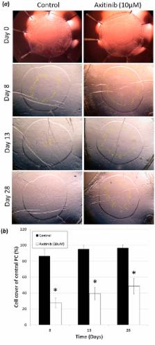
          Secondary visual loss occurs in millions of patients due to a wound-healing response, known as posterior capsule opacification (PCO), following cataract surgery. An intraocular lens (IOL) is implanted into residual lens tissue, known as the capsular bag, following cataract removal. Standard IOLs allow the anterior and posterior capsules to become physically connected. This places pressure on the IOL and improves contact with the underlying posterior capsule. New open bag IOL designs separate the anterior capsule and posterior capsules and further reduce PCO incidence. It is hypothesised that this results from reduced cytokine availability due to greater irrigation of the bag. We therefore explored the role of growth factor restriction on PCO using human lens cell and tissue culture models. We demonstrate that cytokine dilution, by increasing medium volume, significantly reduced cell coverage in both closed and open capsular bag models. This coincided with reduced cell density and myofibroblast formation. A screen of 27 cytokines identified nine candidates whose expression profile correlated with growth. In particular, VEGF was found to regulate cell survival, growth and myofibroblast formation. VEGF provides a therapeutic target to further manage PCO development and will yield best results when used in conjunction with open bag IOL designs.

          Related collections

          Most cited references39

          • Record: found
          • Abstract: found
          • Article: not found

          Intravitreal bevacizumab (Avastin) for neovascular age-related macular degeneration.

          To report the short-term safety, biologic effect, and a possible mechanism of action of intravitreal bevacizumab in patients with neovascular age-related macular degeneration (AMD). Interventional, consecutive, retrospective case series. Eighty-one eyes of 79 patients with subfoveal neovascular AMD. Patients received intravitreal bevacizumab (1.25 mg) on a monthly basis until macular edema, subretinal fluid (SRF), and/or pigment epithelial detachment (PED) resolved. Ophthalmic evaluations included nonstandardized Snellen visual acuity (VA), complete ophthalmic examination, fluorescein angiography, and optical coherence tomography (OCT). Assessments of safety, changes in Snellen VA, OCT retinal thickness, and angiographic lesion characteristics were performed. No significant ocular or systemic side effects were observed. Most patients (55%) had a reduction of >10% of baseline retinal thickness at 1 week after the injection. At 4 weeks after injection, 30 of 81 eyes demonstrated complete resolution of retinal edema, SRF, and PEDs. Of the 51 eyes with 8 weeks' follow-up, 25 had complete resolution of retinal thickening, SRF, and PEDs. At 1, 4, 8,and 12 weeks, the mean retinal thickness of the central 1 mm was decreased by 61, 92, 89, and 67 mum, respectively (P<0.0001 for 1, 4, and 8 weeks and P<0.01 for 12 weeks). At 4 and 8 weeks, mean VA improved from 20/200 to 20/125 (P<0.0001). Median vision improved from 20/200 to 20/80(-) at 4 weeks and from 20/200 to 20/80 at 8 weeks. Short-term results suggest that intravitreal bevacizumab (1.25 mg) is well tolerated and associated with improvement in VA, decreased retinal thickness by OCT, and reduction in angiographic leakage in most patients, the majority of whom had previous treatment with photodynamic therapy and/or pegaptanib. Further evaluation of intravitreal bevacizumab for the treatment of choroidal neovascularization is warranted.
            Bookmark
            • Record: found
            • Abstract: found
            • Article: not found

            Myofibroblasts.

            Boris Hinz (2016)
            Myofibroblasts are activated in response to tissue injury with the primary task to repair lost or damaged extracellular matrix. Enhanced collagen secretion and subsequent contraction - scarring - are part of the normal wound healing response and crucial to restore tissue integrity. Due to myofibroblasts ability to repair but not regenerate, accumulation of scar tissue is always associated with reduced organ performance. This is a fair price to pay by the body for not falling apart. Whereas myofibroblasts typically vanish after successful repair, dysregulation of the normal repair process can lead to persistent myofibroblast activation, for instance by chronic inflammation or mechanical stress in the tissue. Excessive repair leads to the accumulation of stiff collagenous ECM contractures - fibrosis - with dramatic consequences for organ function. The clinical need to terminate detrimental myofibroblast activities has stimulated researchers to answer a number of essential questions: where do myofibroblasts come from, what are the factors leading to their activation, how do we discriminate myofibroblasts from other cells, what is the molecular basis for their contractile activity, and how can we stop or at least control them? This article reviews the current state of the myofibroblast literature by emphasizing their role in ocular repair and fibrosis. It appears that although the eye is quite an extraordinary organ, ocular myofibroblasts behave or misbehave just like their siblings in other organs.
              Bookmark
              • Record: found
              • Abstract: found
              • Article: not found

              Inhibition of PDGF, VEGF and FGF signalling attenuates fibrosis.

              BIBF 1000 is a small molecule inhibitor targeting the receptor kinases of platelet-derived growth factor (PDGF), basic fibroblast growth factor and vascular endothelial growth factor, which have known roles in the pathogenesis of pulmonary fibrosis. The anti-fibrotic potential of BIBF 1000 was determined in a rat model of bleomycin-induced lung fibrosis and in an ex vivo fibroblast differentiation assay. Rats exposed to a single intra-tracheal injection of bleomycin were treated with BIBF 1000 starting 10 days after bleomycin administration. To gauge for anti-fibrotic activity, collagen deposition and pro-fibrotic growth factor gene expression was analysed in isolated lungs. Furthermore, the activity of BIBF 1000 was compared with imatinib mesylate (combined PDGF receptor, c-kit and c-abl kinase inhibitor) and SB-431542 (transforming growth factor (TGF)-beta receptor I kinase inhibitor) in an ex vivo TGF-beta-driven fibroblast to myofibroblast differentiation assay, performed in primary human bronchial fibroblasts. Treatment of rats with BIBF 1000 resulted in the attenuation of fibrosis as assessed by the reduction of collagen deposition and the inhibition of pro-fibrotic gene expression. In the cellular assay both SB-431542 and BIBF 1000 showed dose-dependent inhibition of TGF-beta-induced differentiation, whereas imatinib mesylate was inactive. BIBF 1000, or related small molecules with a similar kinase inhibition profile, may represent a novel therapeutic approach for the treatment of idiopathic pulmonary fibrosis.
                Bookmark

                Author and article information

                Journal
                Sci Rep
                Sci Rep
                Scientific Reports
                Nature Publishing Group
                2045-2322
                14 April 2016
                2016
                : 6
                : 24453
                Affiliations
                [1 ]School of Biological Sciences, University of East Anglia , Norwich NR4 7TJ, UK
                [2 ]King Edward VII Hospital , London, UK
                Author notes
                Article
                srep24453
                10.1038/srep24453
                4831005
                27076230
                10c71614-aae5-4e44-8afd-ac6a659f86c9
                Copyright © 2016, Macmillan Publishers Limited

                This work is licensed under a Creative Commons Attribution 4.0 International License. The images or other third party material in this article are included in the article’s Creative Commons license, unless indicated otherwise in the credit line; if the material is not included under the Creative Commons license, users will need to obtain permission from the license holder to reproduce the material. To view a copy of this license, visit http://creativecommons.org/licenses/by/4.0/

                History
                : 26 October 2015
                : 29 March 2016
                Categories
                Article

                Uncategorized
                Uncategorized

                Comments

                Comment on this article