3
views
0
recommends
+1 Recommend
0 collections
    0
    shares
      • Record: found
      • Abstract: found
      • Article: found
      Is Open Access

      Recognition of Cognitive Impairment in Adult Moyamoya Disease: A Classifier Based on High-Order Resting-State Functional Connectivity Network

      research-article

      Read this article at

      Bookmark
          There is no author summary for this article yet. Authors can add summaries to their articles on ScienceOpen to make them more accessible to a non-specialist audience.

          Abstract

          Objective: Vascular cognitive impairment (VCI) is a common complication in adult patients with moyamoya disease (MMD), and is reversible by surgical revascularization in its early stage of mild VCI. However, accurate diagnosis of mild VCI is difficult based on neuropsychological examination alone. This study proposed a method of dynamic resting-state functional connectivity (FC) network to recognize global cognitive impairment in MMD.

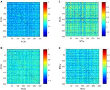
          Methods: For MMD, 36 patients with VCI and 43 patients with intact cognition (Non-VCI) were included, as well as 26 normal controls (NCs). Using resting-state fMRI, dynamic low-order FC networks were first constructed with multiple brain regions which were generated through a sliding window approach and correlated in temporal dimension. In order to obtain more information of network interactions along the time, high-order FC networks were established by calculating correlations among each pair of brain regions. Afterwards, a sparse representation-based classifier was constructed to recognize MMD (experiment 1) and its cognitive impairment (experiment 2) with features extracted from both low- and high-order FC networks. Finally, the ten-fold cross-validation strategy was proposed to train and validate the performance of the classifier.

          Results: The three groups did not differ significantly in demographic features ( p > 0.05), while the VCI group exhibited the lowest MMSE scores ( p = 0.001). The Non-VCI and NCs groups did not differ significantly in MMSE scores ( p = 0.054). As for the classification between MMD and NCs, the area under the receiver operating characteristic curve (AUC), accuracy, sensitivity, and specificity of the classifier reached 90.70, 88.57, 93.67, and 73.08%, respectively. While for the classification between VCI and Non-VCI, the AUC, accuracy, sensitivity, and specificity of the classifier reached 91.02, 84.81, 80.56, and 88.37%, respectively.

          Conclusion: This study not only develops a promising classifier to recognize VCI in adult MMD in its early stage, but also implies the significance of time-varying properties in dynamic FC networks.

          Related collections

          Most cited references38

          • Record: found
          • Abstract: found
          • Article: not found

          Automated anatomical labeling of activations in SPM using a macroscopic anatomical parcellation of the MNI MRI single-subject brain.

          An anatomical parcellation of the spatially normalized single-subject high-resolution T1 volume provided by the Montreal Neurological Institute (MNI) (D. L. Collins et al., 1998, Trans. Med. Imag. 17, 463-468) was performed. The MNI single-subject main sulci were first delineated and further used as landmarks for the 3D definition of 45 anatomical volumes of interest (AVOI) in each hemisphere. This procedure was performed using a dedicated software which allowed a 3D following of the sulci course on the edited brain. Regions of interest were then drawn manually with the same software every 2 mm on the axial slices of the high-resolution MNI single subject. The 90 AVOI were reconstructed and assigned a label. Using this parcellation method, three procedures to perform the automated anatomical labeling of functional studies are proposed: (1) labeling of an extremum defined by a set of coordinates, (2) percentage of voxels belonging to each of the AVOI intersected by a sphere centered by a set of coordinates, and (3) percentage of voxels belonging to each of the AVOI intersected by an activated cluster. An interface with the Statistical Parametric Mapping package (SPM, J. Ashburner and K. J. Friston, 1999, Hum. Brain Mapp. 7, 254-266) is provided as a freeware to researchers of the neuroimaging community. We believe that this tool is an improvement for the macroscopical labeling of activated area compared to labeling assessed using the Talairach atlas brain in which deformations are well known. However, this tool does not alleviate the need for more sophisticated labeling strategies based on anatomical or cytoarchitectonic probabilistic maps.
            Bookmark
            • Record: found
            • Abstract: found
            • Article: not found

            Complex network measures of brain connectivity: uses and interpretations.

            Brain connectivity datasets comprise networks of brain regions connected by anatomical tracts or by functional associations. Complex network analysis-a new multidisciplinary approach to the study of complex systems-aims to characterize these brain networks with a small number of neurobiologically meaningful and easily computable measures. In this article, we discuss construction of brain networks from connectivity data and describe the most commonly used network measures of structural and functional connectivity. We describe measures that variously detect functional integration and segregation, quantify centrality of individual brain regions or pathways, characterize patterns of local anatomical circuitry, and test resilience of networks to insult. We discuss the issues surrounding comparison of structural and functional network connectivity, as well as comparison of networks across subjects. Finally, we describe a Matlab toolbox (http://www.brain-connectivity-toolbox.net) accompanying this article and containing a collection of complex network measures and large-scale neuroanatomical connectivity datasets. Copyright (c) 2009 Elsevier Inc. All rights reserved.
              Bookmark
              • Record: found
              • Abstract: found
              • Article: not found

              Dissociable intrinsic connectivity networks for salience processing and executive control.

              Variations in neural circuitry, inherited or acquired, may underlie important individual differences in thought, feeling, and action patterns. Here, we used task-free connectivity analyses to isolate and characterize two distinct networks typically coactivated during functional MRI tasks. We identified a "salience network," anchored by dorsal anterior cingulate (dACC) and orbital frontoinsular cortices with robust connectivity to subcortical and limbic structures, and an "executive-control network" that links dorsolateral frontal and parietal neocortices. These intrinsic connectivity networks showed dissociable correlations with functions measured outside the scanner. Prescan anxiety ratings correlated with intrinsic functional connectivity of the dACC node of the salience network, but with no region in the executive-control network, whereas executive task performance correlated with lateral parietal nodes of the executive-control network, but with no region in the salience network. Our findings suggest that task-free analysis of intrinsic connectivity networks may help elucidate the neural architectures that support fundamental aspects of human behavior.
                Bookmark

                Author and article information

                Contributors
                Journal
                Front Neural Circuits
                Front Neural Circuits
                Front. Neural Circuits
                Frontiers in Neural Circuits
                Frontiers Media S.A.
                1662-5110
                21 December 2020
                2020
                : 14
                : 603208
                Affiliations
                [1] 1Department of Neurosurgery, Huashan Hospital, Fudan University , Shanghai, China
                [2] 2Department of Electronic Engineering, Fudan University , Shanghai, China
                Author notes

                Edited by: Teppei Matsui, The University of Tokyo, Japan

                Reviewed by: Ken Kazumata, Hokkaido University, Japan; Han Zhang, University of North Carolina at Chapel Hill, United States

                *Correspondence: Yu-Xiang Gu guyuxiang1972@ 123456126.com

                †These authors have contributed equally to this work

                Article
                10.3389/fncir.2020.603208
                7779761
                33408614
                113346aa-339a-4d57-aac6-e9c4f5326834
                Copyright © 2020 Lei, Chen, Su, Zhang, Yang, Gao, Ni, Chen, Yu, Gu and Mao.

                This is an open-access article distributed under the terms of the Creative Commons Attribution License (CC BY). The use, distribution or reproduction in other forums is permitted, provided the original author(s) and the copyright owner(s) are credited and that the original publication in this journal is cited, in accordance with accepted academic practice. No use, distribution or reproduction is permitted which does not comply with these terms.

                History
                : 05 September 2020
                : 03 December 2020
                Page count
                Figures: 7, Tables: 2, Equations: 7, References: 38, Pages: 11, Words: 6074
                Funding
                Funded by: National Natural Science Foundation of China 10.13039/501100001809
                Categories
                Neuroscience
                Original Research

                Neurosciences
                moyamoya disease,resting-state,fmri,vascular cognitive impairment,functional connectivity,sliding window,functional dynamics

                Comments

                Comment on this article