55
views
0
recommends
+1 Recommend
0 collections
    0
    shares
      • Record: found
      • Abstract: found
      • Article: found
      Is Open Access

      Basal Cell Carcinoma Presenting as a Perianal Ulcer and Treated with Radiotherapy

      letter

      Read this article at

      Bookmark
          There is no author summary for this article yet. Authors can add summaries to their articles on ScienceOpen to make them more accessible to a non-specialist audience.

          Abstract

          Dear Editor: Basal cell carcinoma (BCC) in the perianal and genital areas accounts for <1% of all BCCs1. If the patient is unsuitable or unwilling to receive surgical treatment, topical photodynamic therapy and radiotherapy can be considered alternative treatment options2. In this report, we describe a case of BCC that presented as a perianal ulcer and showed a good response to radiotherapy. An 83-year-old man presented with an erythematous ulcer on his perianal area for 3~4 years. The skin lesion was slowly growing, but did not cause pain or bleeding. He did not report any gastrointestinal symptoms. He had a medical history of hypertension, diabetes mellitus, subarachnoid hemorrhage. 
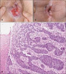
On examination, a single 3.0×3.0 cm-sized, erythematous, asymptomatic ulcer with raised edges was observed on the perianal area (Fig. 1A). Under the clinical impression of Paget disease or Crohn disease, punch biopsy was performed. Histopathologically, nodular masses of basaloid cells extended into the dermis and showed a peripheral palisading pattern, which were consistent with BCC (Fig. 1B). He decided to undergo radiotherapy due to the high operative risk associated with his old age and history of subarachnoid hemorrhage. He received 3 Gy per fraction for 3 times a week for a total dose of 51 Gy over 17 fractions. At the end of radiotherapy, no acute radiogenic skin toxicities such as erythema and desquamation were noted. Two months after radiotherapy, the ulcerative lesion showed considerable clinical improvement (Fig. 1C) and still showed no aggravation after 5 month follow-up. The patient refused to undergo an additional skin biopsy; therefore, he scheduled regular follow-up visits. BCC of non-sun-exposed areas is extremely rare. Among the anogenital BCC, the pubis is the most common, followed by the perianal area, the scrotum, and the penis1. Clinical appearance ranged from erythematous papules to noduloplaques, and ulcers. According to a previous report that reviewed 51 anogenital BCCs, ulcerated lesions were seen in 15 cases (29.4%)1. Perianal ulcerative BCC may initially be misdiagnosed as a benign dermatologic or gastrointestinal disease. Perianal Paget disease, cutaneous metastasis of gastrointestinal malignancy and Crohn disease should be excluded3. Radiotherapy can be a treatment option in elderly patients and those with significant medical comorbidities4. In the present case, poor medical conditions and the location, which made it difficult for complete excision, rendered him unsuitable for surgery. However, patients treated with radiotherapy should be closely followed up because BCC treated with radiotherapy recurs more often than that treated with Mohs micrographic surgery; Rowe et al.5 reported that the 5-year recurrence rate of radiotherapy-treated disease is higher (9.8%) than that of Mohs micrographic surgery (5.6%). Also, the potential for radiogenic toxicity in the skin should not be ignored, especially when it might cause severe functional discomfort. In summary, we describe a rare case of BCC that presented as a perianal ulcer. Dermatologists should consider BCC in the differential diagnosis of a painless, ulcerated lesion on the perianal area. We also suggest considering radiotherapy in cases of BCC in the perianal area when surgical treatment is not possible.

          Related collections

          Most cited references5

          • Record: found
          • Abstract: found
          • Article: not found

          Mohs surgery is the treatment of choice for recurrent (previously treated) basal cell carcinoma.

          We reviewed all studies (since 1945) reporting recurrence rates for treatment of recurrent (previously treated) basal cell carcinomas (BCC) using surgical excision, radiotherapy, cryotherapy, curettage and electrodesiccation, and Mohs micrographic surgery. The 5-year recurrence rate for Mohs micrographic surgery is 5.6%. The recurrence rate for non-Mohs modalities of 19.9% is nearly four times higher. Individual recurrence rates for the non-Mohs modalities are 17.4% for surgical excision, 40.0% for curettage and electrodesiccation, and 9.8% for radiation therapy. There are no studies reporting 5-year data for cryotherapy. However, the recurrence rate is 13.0% for cryotherapy when the follow-up period is less than five years. The data support the following conclusions: (1) Mohs surgery is the treatment of choice for recurrent BCC; (2) if the patient is not a surgical candidate and the lesion is small, radiation therapy is an alternative that offers a better chance for cure than the other non-Mohs modalities; and (3) curettage and electrodesiccation should not be used to treat recurrent basal cell carcinoma.
            Bookmark
            • Record: found
            • Abstract: found
            • Article: not found

            Cutaneous manifestations of gastrointestinal disease: part I.

            Cutaneous findings are not uncommonly a concomitant finding in patients afflicted with gastrointestinal (GI) diseases. The dermatologic manifestations may precede clinically evident GI disease. Part I of this 2-part CME review focuses on dermatologic findings as they relate to hereditary and nonhereditary polyposis disorders and paraneoplastic disorders. A number of hereditary GI disorders have an increased risk of colorectal carcinomas. These disorders include familial adenomatous polyposis, Peutz-Jeghers syndrome, and juvenile polyposis syndrome. Each disease has its own cutaneous signature that aids dermatologists in the early diagnosis and detection of hereditary GI malignancy. These disease processes are associated with particular gene mutations that can be used in screening and to guide additional genetic counseling. In addition, there is a group of hamartomatous syndromes, some of which are associated with phosphatase and tensin homolog (PTEN) gene mutations, which present with concurrent skin findings. These include Cowden syndrome, Bannayan-Riley-Ruvalcaba syndrome, and Cronkhite-Canada syndrome. Finally, paraneoplastic disorders are another subcategory of GI diseases associated with cutaneous manifestations, including malignant acanthosis nigricans, Leser-Trélat sign, tylosis, Plummer-Vinson syndrome, necrolytic migratory erythema, perianal extramammary Paget disease, carcinoid syndrome, paraneoplastic dermatomyositis, and paraneoplastic pemphigus. Each of these disease processes have been shown to be associated with an increased risk of GI malignancy. This underscores the important role of dermatologists in the diagnosis, detection, monitoring, and treatment of these disorders while consulting and interacting with their GI colleagues.
              Bookmark
              • Record: found
              • Abstract: found
              • Article: not found

              Perianal and genital basal cell carcinoma: A clinicopathologic review of 51 cases.

              Basal cell carcinoma (BCC) occurring on non-sun-exposed sites, especially the perianal and genital regions, is very rare. We analyzed the incidence, clinical and pathologic features, and etiologic and prognostic factors of all non-nevoid perianal and genital BCCs diagnosed at our institution within a defined period (January 1985-September 1996). A retrospective review was performed with the use of patient clinical records and dermatopathologic slides. Cutaneous biopsy samples were tested for the presence of human papillomavirus (HPV) by in situ hybridization using biotinylated pan-HPV and serotype-specific (6, 11, 16, 18, 31, 33, 51) probes. Of all non-nevoid BCC syndrome cases, 51 BCCs (0.27%) were located within the perianal and genital regions. The average age of the patients was 73 years. Nine perianal BCCs occurred in men, 6 in women. Ten BCCs occurred in the pubic area, 18 on the vulva, 6 on the scrotum, and 2 on the penis. Three patients had 2 tumor sites. The average size of BCC was 1.95 cm; 29.4% were ulcerated. Seventeen patients (36%) had a history of skin cancer on sun-exposed sites and 10 (21%) had a possibly relevant associated condition. HPV was not detected in the specimens tested. Treatments included wide excision (n = 32), electrodesiccation and curettage (n = 10), Mohs micrographic surgery (n = 8), and carbon dioxide laser (n = 1). Of 30 patients with 5 years' follow-up or longer, 1 recurrence was noted 7 years after wide excision. There were no metastases. BCC of the perianal and genital skin is rare and exhibits clinical and histologic heterogeneity. Advancing age and local trauma may contribute to the pathogenesis of BCC at these sites.
                Bookmark

                Author and article information

                Journal
                Ann Dermatol
                Ann Dermatol
                AD
                Annals of Dermatology
                Korean Dermatological Association; The Korean Society for Investigative Dermatology
                1013-9087
                2005-3894
                April 2015
                24 March 2015
                : 27
                : 2
                : 212-214
                Affiliations
                Department of Dermatology, Ajou University School of Medicine, Suwon, Korea.
                Author notes
                Corresponding author: Sue Kyung Kim, Department of Dermatology, Ajou University Hospital, 164 WorldCup-ro, Yeongtong-gu, Suwon 443-380, Korea. Tel: 82-31-219-5190, Fax: 82-31-219-5189, ksk9167@ 123456lycos.co.kr
                Article
                10.5021/ad.2015.27.2.212
                4377415
                25834365
                13001e81-3d3d-45e6-90fd-4b7d8652b57e
                Copyright © 2015 The Korean Dermatological Association and The Korean Society for Investigative Dermatology

                This is an Open Access article distributed under the terms of the Creative Commons Attribution Non-Commercial License ( http://creativecommons.org/licenses/by-nc/3.0/) which permits unrestricted non-commercial use, distribution, and reproduction in any medium, provided the original work is properly cited.

                History
                : 29 November 2013
                : 22 April 2014
                : 07 May 2014
                Categories
                Letter to the Editor

                Dermatology
                Dermatology

                Comments

                Comment on this article