198
views
0
recommends
+1 Recommend
0 collections
    0
    shares
      • Record: found
      • Abstract: found
      • Article: found
      Is Open Access

      Neurocysticercosis presenting as Millard Gubler syndrome

      case-report

      Read this article at

      Bookmark
          There is no author summary for this article yet. Authors can add summaries to their articles on ScienceOpen to make them more accessible to a non-specialist audience.

          Abstract

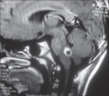
          Neurocysticercosis is a common childhood neurological illness in India. A variety of presentations have been reported in the literature, including weber syndrome. Neurocysticercosis, manifesting as Millard Gubler syndrome, have not been reported in literature. Therefore, we report a child presented to us with Millard Gubler syndrome due to pontomedullary neurocysticercosis and was treated successfully.

          Related collections

          Most cited references8

          • Record: found
          • Abstract: found
          • Article: not found

          Proposed diagnostic criteria for neurocysticercosis.

          Neurocysticercosis is the most common helminthic infection of the CNS but its diagnosis remains difficult. Clinical manifestations are nonspecific, most neuroimaging findings are not pathognomonic, and some serologic tests have low sensitivity and specificity. The authors provide diagnostic criteria for neurocysticercosis based on objective clinical, imaging, immunologic, and epidemiologic data. These include four categories of criteria stratified on the basis of their diagnostic strength, including the following: 1) absolute--histologic demonstration of the parasite from biopsy of a brain or spinal cord lesion, cystic lesions showing the scolex on CT or MRI, and direct visualization of subretinal parasites by funduscopic examination; 2) major--lesions highly suggestive of neurocysticercosis on neuroimaging studies, positive serum enzyme-linked immunoelectrotransfer blot for the detection of anticysticercal antibodies, resolution of intracranial cystic lesions after therapy with albendazole or praziquantel, and spontaneous resolution of small single enhancing lesions; 3) minor--lesions compatible with neurocysticercosis on neuroimaging studies, clinical manifestations suggestive of neurocysticercosis, positive CSF enzyme-linked immunosorbent assay for detection of anticysticercal antibodies or cysticercal antigens, and cysticercosis outside the CNS; and 4) epidemiologic--evidence of a household contact with Taenia solium infection, individuals coming from or living in an area where cysticercosis is endemic, and history of frequent travel to disease-endemic areas. Interpretation of these criteria permits two degrees of diagnostic certainty: 1) definitive diagnosis, in patients who have one absolute criterion or in those who have two major plus one minor and one epidemiologic criterion; and 2) probable diagnosis, in patients who have one major plus two minor criteria, in those who have one major plus one minor and one epidemiologic criterion, and in those who have three minor plus one epidemiologic criterion.
            Bookmark
            • Record: found
            • Abstract: found
            • Article: not found

            Clinical spectrum of 500 children with neurocysticercosis and response to albendazole therapy.

            Neurocysticercosis is a major cause of neurologic illness worldwide. Its manifestations are variable, and somewhat different when it occurs in children. Controversy exists regarding anticysticercal therapy. The clinical, laboratory, and radiographic features of 500 consecutive children with neurocysticercosis were studied; the children were then followed prospectively and their response to albendazole therapy was analyzed. Diagnosis of neurocysticercosis was based primarily on neuroimaging. Computed tomographic (CT) scans, neurocysticercosis serology, chest radiographs, and Mantoux tests were done in all children, and magnetic resonance imaging scans in 10%. All children with multiple lesions, and some randomly allocated children with single, small, enhancing CT lesions received albendazole. CT scans were repeated after 3 to 6 months. There were 272 boys and 228 girls, age range 1 6/12 to 12 6/12 years. Seizures were present in 94.8% of cases; 83.7% had focal seizures. Features of raised intracranial pressure were seen in 30% of patients and focal neurodeficit in 4%. Single lesions were seen in 76% of the children, with perilesional edema in 57.4%. Thirty-four children who had multiple cysts and received albendazole underwent serial CT evaluation. Four showed disappearance of lesions and 22 had reductions in the size or number, to give an overall improvement rate of 76%. Serial CT studies were available on 176 children with single lesions, 90 of whom received albendazole. Improvement (disappearance or reduction in the size of lesions) was observed in 91% (82 of 90) of albendazole-treated children versus 85% (73 of 86) of untreated children. This difference was not significant. No significant side-effects of albendazole were reported. These data indicate that partial seizures and single parenchymal cysts are the most frequent clinical and neuroradiographic manifestations of neurocysticercosis in children. Although albendazole therapy should be considered, especially in children with multiple lesions, many children with isolated neurocysticercosis will improve without antiparasitic therapy.
              Bookmark
              • Record: found
              • Abstract: found
              • Article: not found

              Neurocysticercosis: a review.

              In North America, the largest number of neurosurgical cases stemming from parasitic infections involves the larval form of Taenia solium, the infectious organism causing neurocysticercosis. This infection of the central nervous system (CNS) is most commonly seen in areas with significant immigrant populations and can often present particular challenges to treating physicians. A review of the literature was performed, highlighting the epidemiology, parasitology, and clinical manifestations of neurocysticercosis. Particular attention was paid to 4 locations of neurocysticercosis lesions: parenchymal, subarachnoid/cisternal, intraventricular, and spinal. Both medical and surgical interventions may be necessary for adequate treatment of neurocysticercosis, depending on various factors, including location of lesion. A review of neurocysticercosis, with particular attention paid to location of disease involvement in the CNS, provides important information to the clinical management of this disease.
                Bookmark

                Author and article information

                Journal
                J Neurosci Rural Pract
                J Neurosci Rural Pract
                JNRP
                Journal of Neurosciences in Rural Practice
                Medknow Publications & Media Pvt Ltd (India )
                0976-3147
                0976-3155
                Sep-Dec 2012
                : 3
                : 3
                : 375-377
                Affiliations
                [1] Department of Pediatrics, Institute of Medical Sciences, Banaras Hindu University, Varanasi, Uttar Pradesh, India
                [1 ] Department of Radiodiagnosis, Institute of Medical Sciences, Banaras Hindu University, Varanasi, Uttar Pradesh, India
                Author notes
                Address for correspondence: Dr. Rajniti Prasad, Department of Pediatrics, Institute of Medical Sciences, Banaras Hindu University, Varanasi, Uttar Pradesh, India. E-mail: rajnitip@ 123456gmail.com
                Article
                JNRP-3-375
                10.4103/0976-3147.102634
                3505345
                23189006
                14c10a80-ee99-40a0-8bd2-849af0eeebbf
                Copyright: © Journal of Neurosciences in Rural Practice

                This is an open-access article distributed under the terms of the Creative Commons Attribution-Noncommercial-Share Alike 3.0 Unported, which permits unrestricted use, distribution, and reproduction in any medium, provided the original work is properly cited.

                History
                Categories
                Case Report

                Neurosciences
                neurocysticercosis,cranial nerve palsy,millard gubler syndrome
                Neurosciences
                neurocysticercosis, cranial nerve palsy, millard gubler syndrome

                Comments

                Comment on this article