5
views
0
recommends
+1 Recommend
0 collections
    0
    shares
      • Record: found
      • Abstract: found
      • Article: found
      Is Open Access

      Effect of Sleep Deprivation on the Working Memory-Related N2-P3 Components of the Event-Related Potential Waveform

      research-article

      Read this article at

      Bookmark
          There is no author summary for this article yet. Authors can add summaries to their articles on ScienceOpen to make them more accessible to a non-specialist audience.

          Abstract

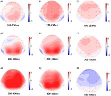
          Working memory is very sensitive to acute sleep deprivation, and many studies focus on the brain areas or network activities of working memory after sleep deprivation. However, little is known about event-related potential (ERP)-related changes in working memory after sleep loss. The purpose of this research was to explore the effects of 36 h of total sleep deprivation (TSD) on working memory through ERPs. Sixteen healthy college students performed working memory tasks while rested and after 36 h of TSD, and electroencephalography (EEG) data were simultaneously recorded while the subjects completed working memory tasks that included different types of stimulus materials. ERP data were statistically analyzed using repeated measurements analysis of variance to observe the changes in the working memory-related N2-P3 components. Compared with baseline before TSD, the amplitude of N2-P3 components related to working memory decreased, and the latency was prolonged after TSD. However, the increased amplitude of the P2 wave and the prolonged latency were found after 36 h of TSD. Thus, TSD can impair working memory capacity, which is characterized by lower amplitude and prolonged latency.

          Related collections

          Most cited references42

          • Record: found
          • Abstract: found
          • Article: not found

          The prefrontal cortex in sleep.

          Experimental data indicate a role for the prefrontal cortex in mediating normal sleep physiology, dreaming and sleep-deprivation phenomena. During nonrandom-eye-movement (NREM) sleep, frontal cortical activity is characterized by the highest voltage and the slowest brain waves compared to other cortical regions. The differences between the self-awareness experienced in waking and its diminution in dreaming can be explained by deactivation of the dorsolateral prefrontal cortex during REM sleep. Here, we propose that this deactivation results from a direct inhibition of the dorsolateral prefrontal cortical neurons by acetylcholine, the release of which is enhanced during REM sleep. Sleep deprivation influences frontal executive functions in particular, which further emphasizes the sensitivity of the prefrontal cortex to sleep.
            Bookmark
            • Record: found
            • Abstract: found
            • Article: not found

            The neural basis of interindividual variability in inhibitory efficiency after sleep deprivation.

            Sleep deprivation results in the loss of our ability to suppress a prepotent response. The extent of decline in this executive function varies across individuals. Here, we used functional magnetic resonance imaging to study the neural correlates of sleep deprivation-induced differences in inhibitory efficiency. Participants performed a go/no-go task after normal sleep and after 24 h of total sleep deprivation. Regardless of the extent of change in inhibitory efficiency, sleep deprivation lowered go/no-go sustained, task-related activation of the ventral and anterior prefrontal (PFC) regions bilaterally. However, individuals better able to maintain inhibitory efficiency after sleep deprivation could be distinguished by lower stop-related, phasic activation of the right ventral PFC during rested wakefulness. These persons also showed a larger rise in such activation both here and in the right insula after sleep deprivation relative to those whose inhibitory efficiency declined.
              Bookmark
              • Record: found
              • Abstract: found
              • Article: not found

              Frontal lobe function, sleep loss and fragmented sleep.

              Recent experimental studies involving total sleep loss, sleep reduction and clinically related sleep fragmentation report impaired performance on tasks of frontal lobe or executive function, including measures of verbal fluency, creativity and planning skills. Severity of sleep disturbance in obstructive sleep apnoea syndrome (OSAS) is correlated with level of executive impairment, with some residual impairment despite treatment (continuous positive airway pressure - CPAP). Executive impairment appears to be more closely related to hypoxaemic events rather than daytime sleepiness. Studies of electroencephalographic (EEG) changes throughout the course of sleep and following sleep deprivation as well as functional neuroimaging and psychophysiological changes (event-related potentials - ERPs) following sleep deprivation provide further indication of the relative importance of the frontal regions of the brain to sleep. However, neurocognitive studies present many inconsistencies, task classification is often ambiguous and, in the absence of any unifying explanation at the level of cognitive mechanisms, the overall picture is one of a disparate range of impairment following sleep loss and sleep fragmentation. Poorly defined concepts of frontal lobe function, executive function, memory and attention, using tasks largely developed with more severe deficit levels in mind, create further difficulties in interpreting current findings.
                Bookmark

                Author and article information

                Contributors
                Journal
                Front Neurosci
                Front Neurosci
                Front. Neurosci.
                Frontiers in Neuroscience
                Frontiers Media S.A.
                1662-4548
                1662-453X
                19 May 2020
                2020
                : 14
                : 469
                Affiliations
                [1] 1School of Psychology, Beijing Sport University , Beijing, China
                [2] 2Institute of Psychology, Chinese Academy of Sciences , Beijing, China
                [3] 3Naval Special Forces Recuperation Center , Qingdao, China
                Author notes

                Edited by: Ritchie Edward Brown, VA Boston Healthcare System, United States

                Reviewed by: Jaime Tartar, Nova Southeastern University, United States; Hiromasa Funato, Toho University, Japan

                *Correspondence: Yongcong Shao, budeshao@ 123456aliyun.com

                This article was submitted to Sleep and Circadian Rhythms, a section of the journal Frontiers in Neuroscience

                Article
                10.3389/fnins.2020.00469
                7248549
                32508572
                14ccda23-f311-4878-b0fe-3cc2b2446715
                Copyright © 2020 Peng, Dai, Ba, Zhang, Shao and Tian.

                This is an open-access article distributed under the terms of the Creative Commons Attribution License (CC BY). The use, distribution or reproduction in other forums is permitted, provided the original author(s) and the copyright owner(s) are credited and that the original publication in this journal is cited, in accordance with accepted academic practice. No use, distribution or reproduction is permitted which does not comply with these terms.

                History
                : 12 February 2020
                : 15 April 2020
                Page count
                Figures: 5, Tables: 3, Equations: 0, References: 51, Pages: 9, Words: 0
                Categories
                Neuroscience
                Original Research

                Neurosciences
                sleep deprivation,working memory,event related potentials,electroencephalography,n-back

                Comments

                Comment on this article