25
views
0
recommends
+1 Recommend
0 collections
    0
    shares
      • Record: found
      • Abstract: found
      • Article: found
      Is Open Access

      The Fanconi Anemia DNA Repair Pathway Is Regulated by an Interaction between Ubiquitin and the E2-like Fold Domain of FANCL*

      research-article

      Read this article at

      Bookmark
          There is no author summary for this article yet. Authors can add summaries to their articles on ScienceOpen to make them more accessible to a non-specialist audience.

          Abstract

          Background: The E3 ligase FANCL monoubiquitinates FANCD2 in a critical step in the repair of DNA interstrand crosslinks.

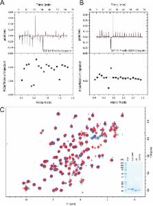
          Results: FANCL binds ubiquitin non-covalently via its N-terminal E2-like fold.

          Conclusion: Monoubiquitination of FANCD2 is regulated via a non-covalent interaction between FANCL and ubiquitin.

          Significance: This interaction represents an additional layer of regulation of the Fanconi Anemia pathway, and a targetable interface.

          Abstract

          The Fanconi Anemia (FA) DNA repair pathway is essential for the recognition and repair of DNA interstrand crosslinks (ICL). Inefficient repair of these ICL can lead to leukemia and bone marrow failure. A critical step in the pathway is the monoubiquitination of FANCD2 by the RING E3 ligase FANCL. FANCL comprises 3 domains, a RING domain that interacts with E2 conjugating enzymes, a central domain required for substrate interaction, and an N-terminal E2-like fold (ELF) domain. The ELF domain is found in all FANCL homologues, yet the function of the domain remains unknown. We report here that the ELF domain of FANCL is required to mediate a non-covalent interaction between FANCL and ubiquitin. The interaction involves the canonical Ile44 patch on ubiquitin, and a functionally conserved patch on FANCL. We show that the interaction is not necessary for the recognition of the core complex, it does not enhance the interaction between FANCL and Ube2T, and is not required for FANCD2 monoubiquitination in vitro. However, we demonstrate that the ELF domain is required to promote efficient DNA damage-induced FANCD2 monoubiquitination in vertebrate cells, suggesting an important function of ubiquitin binding by FANCL in vivo.

          Related collections

          Most cited references38

          • Record: found
          • Abstract: found
          • Article: not found

          RAD6-dependent DNA repair is linked to modification of PCNA by ubiquitin and SUMO.

          The RAD6 pathway is central to post-replicative DNA repair in eukaryotic cells; however, the machinery and its regulation remain poorly understood. Two principal elements of this pathway are the ubiquitin-conjugating enzymes RAD6 and the MMS2-UBC13 heterodimer, which are recruited to chromatin by the RING-finger proteins RAD18 and RAD5, respectively. Here we show that UBC9, a small ubiquitin-related modifier (SUMO)-conjugating enzyme, is also affiliated with this pathway and that proliferating cell nuclear antigen (PCNA) -- a DNA-polymerase sliding clamp involved in DNA synthesis and repair -- is a substrate. PCNA is mono-ubiquitinated through RAD6 and RAD18, modified by lysine-63-linked multi-ubiquitination--which additionally requires MMS2, UBC13 and RAD5--and is conjugated to SUMO by UBC9. All three modifications affect the same lysine residue of PCNA, suggesting that they label PCNA for alternative functions. We demonstrate that these modifications differentially affect resistance to DNA damage, and that damage-induced PCNA ubiquitination is elementary for DNA repair and occurs at the same conserved residue in yeast and humans.
            Bookmark
            • Record: found
            • Abstract: found
            • Article: not found

            Ubiquitin-binding domains - from structures to functions.

            Ubiquitin-binding domains (UBDs) are modular elements that bind non-covalently to the protein modifier ubiquitin. Recent atomic-level resolution structures of ubiquitin-UBD complexes have revealed some of the mechanisms that underlie the versatile functions of ubiquitin in vivo. The preferences of UBDs for ubiquitin chains of specific length and linkage are central to these functions. These preferences originate from multimeric interactions, whereby UBDs synergistically bind multiple ubiquitin molecules, and from contacts with regions that link ubiquitin molecules into a polymer. The sequence context of UBDs and the conformational changes that follow their binding to ubiquitin also contribute to ubiquitin signalling. These new structure-based insights provide strategies for controlling cellular processes by targeting ubiquitin-UBD interfaces.
              Bookmark
              • Record: found
              • Abstract: found
              • Article: not found

              Ubiquitin-binding proteins: decoders of ubiquitin-mediated cellular functions.

              Ubiquitin acts as a versatile cellular signal that controls a wide range of biological processes including protein degradation, DNA repair, endocytosis, autophagy, transcription, immunity, and inflammation. The specificity of ubiquitin signaling is achieved by alternative conjugation signals (monoubiquitin and ubiquitin chains) and interactions with ubiquitin-binding proteins (known as ubiquitin receptors) that decode ubiquitinated target signals into biochemical cascades in the cell. Herein, we review the current knowledge pertaining to the structural and functional features of ubiquitin-binding proteins and the mechanisms by which they recognize various types of ubiquitin topologies. The combinatorial use of diverse ubiquitin-binding domains (UBDs) in full-length proteins, selective recognition of chains with distinct linkages and length, and posttranslational modifications of ubiquitin receptors or multivalent interactions within protein complexes illustrate a few mechanisms by which a circuitry of signaling networks can be rewired by ubiquitin-binding proteins to control cellular functions in vivo.
                Bookmark

                Author and article information

                Journal
                J Biol Chem
                J. Biol. Chem
                jbc
                jbc
                JBC
                The Journal of Biological Chemistry
                American Society for Biochemistry and Molecular Biology (11200 Rockville Pike, Suite 302, Rockville, MD 20852-3110, U.S.A. )
                0021-9258
                1083-351X
                21 August 2015
                6 July 2015
                6 July 2015
                : 290
                : 34
                : 20995-21006
                Affiliations
                From the []Protein Structure and Function Laboratory, Lincoln's Inn Fields Laboratories of the London Research Institute, Cancer Research, United Kingdom, 44 Lincoln's Inn Fields, London WC2A 3LY, United Kingdom,
                [§ ]Medical Research Council Protein Phosphorylation and Ubiquitylation Unit, College of Life Sciences and
                []Scottish Institute for Cell Signalling, College of Life Sciences, University of Dundee, Dow Street, Dundee DD1 5EH, United Kingdom, and
                []Protein Science Group, School of Biosciences, University of Kent, Canterbury, Kent CT2 7NZ, United Kingdom
                Author notes
                [1 ] To whom correspondence may be addressed: Medical Research Council Protein Phosphorylation and Ubiquitylation Unit, College of Life Sciences, University of Dundee, Dow Street, Dundee DD1 5EH, UK. E-mail: A.F.Alpi@ 123456dundee.ac.uk .
                [2 ] To whom correspondence may be addressed: Medical Research Council Protein Phosphorylation and Ubiquitylation Unit, College of Life Sciences, University of Dundee, Dow Street, Dundee DD1 5EH, UK. E-mail: h.walden@ 123456dundee.ac.uk .
                Article
                M115.675835
                10.1074/jbc.M115.675835
                4543658
                26149689
                16b8d7ad-c24b-406a-a66a-f75cedb7d460
                © 2015 by The American Society for Biochemistry and Molecular Biology, Inc.

                Author's Choice—Final version free via Creative Commons CC-BY license.

                History
                : 2 July 2015
                Categories
                Protein Structure and Folding

                Biochemistry
                dna repair,e3 ubiquitin ligase,isothermal titration calorimetry (itc),nuclear magnetic resonance (nmr),ubiquitin,structure

                Comments

                Comment on this article