3
views
0
recommends
+1 Recommend
0 collections
    0
    shares
      • Record: found
      • Abstract: found
      • Article: found
      Is Open Access

      Cost-effective modified endoscopic vacuum therapy for the treatment of gastrointestinal transmural defects: step-by-step process of manufacturing and its advantages

      brief-report

      Read this article at

      Bookmark
          There is no author summary for this article yet. Authors can add summaries to their articles on ScienceOpen to make them more accessible to a non-specialist audience.

          Video

          Video 1

          Cost-effective modified endoscopic vacuum therapy for GI transmural defects. Step-by-step process of manufacturing and potential advantages.

          • 1.

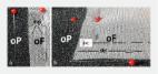
            Cut half gauze to the ideal size to cover only the fenestrated portion of the nasogastric tube (NGT).

          • 2.

            Wrap the gauze around the fenestrated portion of the NGT. The assistance of another person is important in this process.

          • 3.

            Cut the antimicrobial incise drape to match the size of the fenestrated portion of the NGT. Note that the incise drape is a very strong adhesive; therefore, 3 people are usually required to assemble it properly.

          • 4.

            Next, the suture is used to fix the gauze and drape to the NGT. Perform fixation of the modified sponge in 3 places. The first knot is in the proximal portion, just below the last fenestra of the NGT, as a marker of where the vacuum system starts. The second knot is at the distal end, to avoid migration of the modified sponge. The third knot is in the middle of the modified sponge, which is essential to serve as a guide during endoscopic placement. For example, in cases of defects without collection in which the sponge will be placed in an intraluminal position, it is ideal to place the vacuum system in the middle of the defect; in cases of intracavitary placement, it will work as a guide to how much of the modified sponge will be inside the collection.

          • 5.

            Finally, use a needle to make innumerable punctures in the modified sponge system to obtain adequate aspiration. An 18G needle is recommended because, in addition to having an adequate diameter, it is very sharp, which facilitates perforation of the modified sponge system.

          • 6.

            After creation of the modified endoscopic vacuum therapy, the functionality test is performed. Turn on the wall suction system, connect the distal end of the NGT to the tube of the canister connected on the wall, and place the NGT inside a bowl with a liquid solution. The aspiration of a large amount of liquid indicates proper functioning of the modified endoscopic vacuum therapy system.

          • 7.

            The device is then ready to be positioned endoscopically in the patient. After proper positioning, connect the NGT to the suction tube to avoid migration of the device upon removal of the scope.

          • 8.

            In addition to the cost-effective device as described, in our practice we also use wall suction to reduce costs associated with the use of the vacuum machine.

          • 9.

            Use the antimicrobial incise drape to seal the connection between the NGT and the suction tube to avoid leakage within the connection.

          • 10.

            Last, owing to instability of the negative wall pressure, a 20F intravenous catheter is connected to the tube to maintain a negative pressure between –75 and –150 mmHg, as confirmed by laboratory studies performed by our group.

          Related collections

          Most cited references21

          • Record: found
          • Abstract: found
          • Article: not found

          Endoscopic vacuum therapy for various defects of the upper gastrointestinal tract.

          Postoperative, iatrogenic or spontaneous upper gastrointestinal defects result in significant morbidity and mortality of the patients. In the last few years, endoscopic vacuum therapy (EVT) has been recognized as a new promising method for repairing upper gastrointestinal defects of different etiology. However, probably due to insufficient data and no commercially available system for EVT of the upper gastrointestinal tract, until the end of 2014, covering of esophageal defects with self-expanding metal stents (SEMS) were still the mainstay of endoscopic therapy. The aim of this article is to review the data available about EVT for various upper gastrointestinal defects.
            Bookmark
            • Record: found
            • Abstract: found
            • Article: not found

            Role of endoscopic vacuum therapy in the management of gastrointestinal transmural defects

            A gastrointestinal (GI) transmural defect is defined as total rupture of the GI wall, and these defects can be divided into three categories: perforations, leaks, and fistulas. Surgical management of these defects is usually challenging and may be associated with high morbidity and mortality rates. Recently, several novel endoscopic techniques have been developed, and endoscopy has become a first-line approach for therapy of these conditions. The use of endoscopic vacuum therapy (EVT) is increasing with favorable results. This technique involves endoscopic placement of a sponge connected to a nasogastric tube into the defect cavity or lumen. This promotes healing via five mechanisms, including macrodeformation, microdeformation, changes in perfusion, exudate control, and bacterial clearance, which is similar to the mechanisms in which skin wounds are treated with commonly employed wound vacuums. EVT can be used in the upper GI tract, small bowel, biliopancreatic regions, and lower GI tract, with variable success rates and a satisfactory safety profile. In this article, we review and discuss the mechanism of action, materials, techniques, efficacy, and safety of EVT in the management of patients with GI transmural defects.
              Bookmark
              • Record: found
              • Abstract: found
              • Article: found
              Is Open Access

              Open-pore film drainage (OFD): a new multipurpose tool for endoscopic negative pressure therapy (ENPT)

              Background and study aims  Endoscopic negative pressure therapy (ENPT) has been developed to treat gastrointestinal leakages. Up to now, ENPT has usually been performed with open-pore polyurethane foam drains (OPD). A big disadvantage of the OPDs is their large diameter. We have developed a new, small-bore open-pore film drainage (OFD). Herein we report our first experience in a case series of 16 patients. Patients and methods  OFD is constructed with a drainage tube and a very thin double-layered open-pore drainage film (Suprasorb CNP, Drainage Film, Lohmann & Rauscher International, Germany). The distal end of the tube is wrapped with only one layer of film. OFD is placed into the gastrointestinal leakage site with common endoscopic techniques. The tube is connected to an electronic vacuum device and continuous negative pressure of –125 mmHg applied. Results  From 2013 to 2016, 16 patients were treated with the new OFD device. In 10 patients, transmural intestinal defects (4 esophageal, 4 rectum/colon, 1 duodenal, 1 pancreatic cyst) were closed with ENPT in median time of 12 days (range 3 – 34 days). Five of the 10 patients were treated solely with OFD devices. In five patients ENPT started with ODP and changed to OFD when the cavity was shrunken to a channel with a small opening. In four patients postoperative gastric reflux was eliminated for 5 to 16 days. Conclusions  Small-bore OFD opens up promising new treatment options within ENPT. OFD can be used in endoscopic closure management of intestinal leakages in the upper and lower gastrointestinal tract. Gastric reflux can be eliminated in an active manner. OFD can be inserted nasally. OFD may be an adequate substitute for OPD, especially when placement of the larger OPD is difficult.
                Bookmark

                Author and article information

                Journal
                VideoGIE
                VideoGIE
                VideoGIE
                Elsevier
                2468-4481
                04 September 2021
                December 2021
                04 September 2021
                : 6
                : 12
                : 523-528
                Affiliations
                [1 ]Gastrointestinal Endoscopy Unit, Gastroenterology Department, Hospital das Clínicas da Faculdade de Medicina da Universidade de São Paulo, São Paulo, Brazil
                [2 ]Division of Gastroenterology, Hepatology, and Endoscopy, Harvard Medical School, Brigham and Women's Hospital, Boston, Massachusetts
                [3 ]Surgery Division, Federal University of Rio Grande do Norte, Natal, Brazil
                Article
                S2468-4481(21)00155-7
                10.1016/j.vgie.2021.08.002
                8645785
                34917860
                1d68254e-a03e-4fdd-83df-40537b7425f0
                © 2021 American Society for Gastrointestinal Endoscopy. Published by Elsevier Inc.

                This is an open access article under the CC BY-NC-ND license (http://creativecommons.org/licenses/by-nc-nd/4.0/).

                History
                Categories
                Tools and Techniques

                evt, endoscopic vacuum therapy,ngt, nasogastric tube,sems, self-expandable metal stent

                Comments

                Comment on this article