14
views
0
recommends
+1 Recommend
0 collections
    0
    shares
      • Record: found
      • Abstract: found
      • Article: found
      Is Open Access

      Corneal Endothelial Cell Density and Morphology in Healthy Egyptian Eyes

      research-article

      Read this article at

      Bookmark
          There is no author summary for this article yet. Authors can add summaries to their articles on ScienceOpen to make them more accessible to a non-specialist audience.

          Abstract

          Purpose

          To evaluate the corneal endothelial cell density and morphology in normal Egyptian eyes.

          Methods

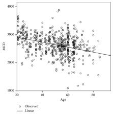
          In total, 568 healthy eyes of 568 Egyptian volunteers aged 20 to 85 years were examined using noncontact specular microscopy for the central corneal thickness (CCT), mean endothelial cell density (MCD), coefficient of variation (CV) in cell area, mean cell area (MCA), and hexagonal cell (Hex) percentage. Variables were compared between sexes and between different age groups.

          Results

          The mean CCT, MCD, and MCA were 514.45 ± 43.04  μm, 2647.50 ± 382.62 cells/mm 2, and 390.59 ± 149.94  μm 2, respectively. MCD and MCA showed no significant differences between men and women ( P=0.171 and 0.099, respectively), whereas CV (%) and Hex (%) showed significant differences ( P=0.024 and 0.015, respectively). CCT ( P=0.007, r = −0.113) and MCD ( P < 0.001, r = −0.357) exhibited a significant negative correlation with age, whereas CV (%) ( P < 0.001, r = 0.341) and MCA ( P=0.008, r = 0.111) exhibited a significant positive correlation. The mean rate of endothelial cell loss from 20 to 85 years of age was 0.3% per year.

          Conclusions

          Our results provide normative data for the corneal endothelium in healthy Egyptian eyes, thus increasing the knowledge base for corneal endothelial cell parameters in healthy Egyptian eyes. Furthermore, our findings can be used as baseline values for comparisons between Egyptian and other populations and for studies of the endothelial cell reserve and capacity for intraocular surgery and corneal transplantation.

          Related collections

          Most cited references35

          • Record: found
          • Abstract: found
          • Article: not found

          Review of corneal endothelial specular microscopy for FDA clinical trials of refractive procedures, surgical devices, and new intraocular drugs and solutions.

          Specular microscopy can provide a noninvasive morphologic analysis of the corneal endothelial cell layer from subjects enrolled in clinical trials. The analysis provides a measure of the endothelial cell physiologic reserve from aging, ocular surgical procedures, pharmaceutical exposure, and general health of the corneal endothelium. The purpose of this review is to discuss normal and stressed endothelial cell morphology, the techniques for determining the morphology parameters, and clinical trial applications.
            Bookmark
            • Record: found
            • Abstract: found
            • Article: not found

            The corneal endothelium.

            The endothelium is a monolayer of cells on the posterior corneal surface that transports water from the stroma into the anterior chamber. This movement of water counters a natural tendency for the stroma to swell and is necessary to maintain a transparent cornea. Embryologic studies, in particular the demonstration of the derivation of the endothelium from the neural crest, have provided insight into the factors that govern the response of this tissue to disease. In some species the endothelium can regenerate after injury, but in man cellular enlargement is the main mechanism of repair after cell loss. A clinical estimate of endothelial cell density and function is provided by specular microscopy, fluorophotometry and pachymetry. In this paper we review the development, structure and function of the corneal endothelium, and then consider the pathological processes that can affect this tissue.
              Bookmark
              • Record: found
              • Abstract: found
              • Article: not found

              Age-related differences in the normal human cornea: a laser scanning in vivo confocal microscopy study.

              To quantify and establish baseline normative data for age-related differences in cellular and innervation density in the normal, healthy, human cornea using laser scanning in vivo confocal microscopy.
                Bookmark

                Author and article information

                Contributors
                Journal
                J Ophthalmol
                J Ophthalmol
                JOPH
                Journal of Ophthalmology
                Hindawi
                2090-004X
                2090-0058
                2019
                24 February 2019
                : 2019
                : 6370241
                Affiliations
                1Ophthalmology Department, Sohag University, Sohag, Egypt
                2Egyptian Police Hospital, Sohag University, Sohag, Egypt
                Author notes

                Academic Editor: Antonio Queiros

                Author information
                http://orcid.org/0000-0003-0261-6856
                http://orcid.org/0000-0002-9662-3602
                http://orcid.org/0000-0002-7286-6124
                http://orcid.org/0000-0002-5731-1972
                http://orcid.org/0000-0002-4355-4973
                http://orcid.org/0000-0001-6436-5576
                Article
                10.1155/2019/6370241
                6409007
                30918718
                1db77af3-7c19-42fd-b46f-b6456a684442
                Copyright © 2019 Marwa Mahmoud Abdellah et al.

                This is an open access article distributed under the Creative Commons Attribution License, which permits unrestricted use, distribution, and reproduction in any medium, provided the original work is properly cited.

                History
                : 18 August 2018
                : 22 December 2018
                : 10 February 2019
                Categories
                Research Article

                Ophthalmology & Optometry
                Ophthalmology & Optometry

                Comments

                Comment on this article