11
views
0
recommends
+1 Recommend
0 collections
    0
    shares
      • Record: found
      • Abstract: found
      • Article: found
      Is Open Access

      Mannitol induces selective astroglial death in the CA1 region of the rat hippocampus following status epilepticus

      research-article

      Read this article at

      Bookmark
          There is no author summary for this article yet. Authors can add summaries to their articles on ScienceOpen to make them more accessible to a non-specialist audience.

          Abstract

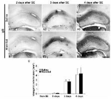
          In the present study, we addressed the question of whether treatment with mannitol, an osmotic diuretic, affects astrogliovascular responses to status epilepticus (SE). In saline-treated animals, astrocytes exhibited reactive astrogliosis in the CA1-3 regions 2-4 days after SE. In the mannitol-treated animals, a large astroglial empty zone was observed in the CA1 region 2 days after SE. This astroglial loss was unrelated to vasogenic edema formation. There was no difference in SE-induced neuronal loss between saline- and mannitol-treated animals. Furthermore, mannitol treatment did not affect astroglial loss and vasogenic edema formation in the dentate gyrus and the piriform cortex. These findings suggest that mannitol treatment induces selective astroglial loss in the CA1 region independent of vasogenic edema formation following SE. These findings support the hypothesis that the susceptibility of astrocytes to SE is most likely due to the distinctive heterogeneity of astrocytes independent of hemodynamics. [BMB Reports 2015; 48(9): 507-512]

          Related collections

          Most cited references27

          • Record: found
          • Abstract: found
          • Article: not found

          Edema and brain trauma.

          Brain edema leading to an expansion of brain volume has a crucial impact on morbidity and mortality following traumatic brain injury (TBI) as it increases intracranial pressure, impairs cerebral perfusion and oxygenation, and contributes to additional ischemic injuries. Classically, two major types of traumatic brain edema exist: "vasogenic" due to blood-brain barrier (BBB) disruption resulting in extracellular water accumulation and "cytotoxic/cellular" due to sustained intracellular water collection. A third type, "osmotic" brain edema is caused by osmotic imbalances between blood and tissue. Rarely after TBI do we encounter a "hydrocephalic edema/interstitial" brain edema related to an obstruction of cerebrospinal fluid outflow. Following TBI, various mediators are released which enhance vasogenic and/or cytotoxic brain edema. These include glutamate, lactate, H(+), K(+), Ca(2+), nitric oxide, arachidonic acid and its metabolites, free oxygen radicals, histamine, and kinins. Thus, avoiding cerebral anaerobic metabolism and acidosis is beneficial to control lactate and H(+), but no compound inhibiting mediators/mediator channels showed beneficial results in conducted clinical trials, despite successful experimental studies. Hence, anti-edematous therapy in TBI patients is still symptomatic and rather non-specific (e.g. mannitol infusion, controlled hyperventilation). For many years, vasogenic brain edema was accepted as the prevalent edema type following TBI. The development of mechanical TBI models ("weight drop," "fluid percussion injury," and "controlled cortical impact injury") and the use of magnetic resonance imaging, however, revealed that "cytotoxic" edema is of decisive pathophysiological importance following TBI as it develops early and persists while BBB integrity is gradually restored. These findings suggest that cytotoxic and vasogenic brain edema are two entities which can be targeted simultaneously or according to their temporal prevalence.
            Bookmark
            • Record: found
            • Abstract: found
            • Article: found
            Is Open Access

            Molecular biology of the blood-brain and the blood-cerebrospinal fluid barriers: similarities and differences

            Efficient processing of information by the central nervous system (CNS) represents an important evolutionary advantage. Thus, homeostatic mechanisms have developed that provide appropriate circumstances for neuronal signaling, including a highly controlled and stable microenvironment. To provide such a milieu for neurons, extracellular fluids of the CNS are separated from the changeable environment of blood at three major interfaces: at the brain capillaries by the blood-brain barrier (BBB), which is localized at the level of the endothelial cells and separates brain interstitial fluid (ISF) from blood; at the epithelial layer of four choroid plexuses, the blood-cerebrospinal fluid (CSF) barrier (BCSFB), which separates CSF from the CP ISF, and at the arachnoid barrier. The two barriers that represent the largest interface between blood and brain extracellular fluids, the BBB and the BCSFB, prevent the free paracellular diffusion of polar molecules by complex morphological features, including tight junctions (TJs) that interconnect the endothelial and epithelial cells, respectively. The first part of this review focuses on the molecular biology of TJs and adherens junctions in the brain capillary endothelial cells and in the CP epithelial cells. However, normal function of the CNS depends on a constant supply of essential molecules, like glucose and amino acids from the blood, exchange of electrolytes between brain extracellular fluids and blood, as well as on efficient removal of metabolic waste products and excess neurotransmitters from the brain ISF. Therefore, a number of specific transport proteins are expressed in brain capillary endothelial cells and CP epithelial cells that provide transport of nutrients and ions into the CNS and removal of waste products and ions from the CSF. The second part of this review concentrates on the molecular biology of various solute carrier (SLC) transport proteins at those two barriers and underlines differences in their expression between the two barriers. Also, many blood-borne molecules and xenobiotics can diffuse into brain ISF and then into neuronal membranes due to their physicochemical properties. Entry of these compounds could be detrimental for neural transmission and signalling. Thus, BBB and BCSFB express transport proteins that actively restrict entry of lipophilic and amphipathic substances from blood and/or remove those molecules from the brain extracellular fluids. The third part of this review concentrates on the molecular biology of ATP-binding cassette (ABC)-transporters and those SLC transporters that are involved in efflux transport of xenobiotics, their expression at the BBB and BCSFB and differences in expression in the two major blood-brain interfaces. In addition, transport and diffusion of ions by the BBB and CP epithelium are involved in the formation of fluid, the ISF and CSF, respectively, so the last part of this review discusses molecular biology of ion transporters/exchangers and ion channels in the brain endothelial and CP epithelial cells.
              Bookmark
              • Record: found
              • Abstract: found
              • Article: not found

              Lesion genesis in a subset of patients with multiple sclerosis: a role for innate immunity?

              Lesions obtained early in the course of multiple sclerosis (MS) have been studied immunocytochemically, and compared with the early stages of the experimental lesion induced in rats by the intraspinal injection of lipopolysaccharide. Large hemispheric or double hemispheric sections were examined from patients who had died in the course of acute or early relapsing multiple sclerosis. In MS patients exhibiting hypoxia-like lesions [Pattern III; Lucchinetti et al. Ann Neurol (2000) 47: 707-17], focal areas in the white matter showed mild oedema, microglial activation and mild axonal injury in the absence of overt demyelination. In such lesions T-cell infiltration was mild and restricted to the perivascular space. Myeloperoxidase and the inducible form of nitric oxide synthase were expressed primarily by microglia, and the activated form of these cells was associated with extracellular deposition of precipitated fibrin. In addition, these lesions showed up-regulation of proteins involved in tissue preconditioning. When active demyelination started, lesions were associated with massive T-cell infiltration and microglia and macrophages expressed all activation markers studied. Similar tissue alterations were found in rats in the pre-demyelinating stage of lesions induced by the focal injection of bacterial lipopolysaccharide into the spinal white matter. We suggest that the areas of microglial activation represent an early stage of tissue injury, which precedes the formation of hypoxia-like demyelinated plaques. The findings indicate that mechanisms associated with innate immunity may play a role in the formation of hypoxia-like demyelinating lesions in MS.
                Bookmark

                Author and article information

                Journal
                BMB Rep
                BMB Rep
                ksbmb
                BMB Reports
                Korean Society for Biochemistry and Molecular Biology
                1976-6696
                1976-670X
                September 2015
                : 48
                : 9
                : 507-512
                Affiliations
                Department of Anatomy and Neurobiology, Institute of Epilepsy Research, College of Medicine, Hallym University, Chunchon 24252, Korea
                Author notes
                [* ]Tel: +82-33-248-2524; Fax: +82-33-248-2525; E-mail: tckang@ 123456hallym.ac.kr
                Article
                BMB-48-507
                10.5483/BMBRep.2015.48.9.013
                4641234
                25703536
                1f465c1d-59a6-4b03-a58f-1f6aad0c3d96
                Copyright © 2015, Korean Society for Biochemistry and Molecular Biology

                This is an open-access article distributed under the terms of the Creative Commons Attribution Non-Commercial License ( http://creativecommons.org/licenses/by-nc/3.0) which permits unrestricted non-commercial use, distribution, and reproduction in any medium, provided the original work is properly cited.

                History
                : 22 January 2015
                : 10 February 2015
                : 16 February 2015
                Categories
                Research Article

                astroglial death,blood-brain barrier,epilepsy,mannitol,status epilepticus

                Comments

                Comment on this article