- Record: found
- Abstract: found
- Article: found
A tissue-bioengineering strategy for modeling rare human kidney diseases in vivo
Read this article at
Abstract
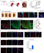
The lack of animal models for some human diseases precludes our understanding of disease mechanisms and our ability to test prospective therapies in vivo. Generation of kidney organoids from Tuberous Sclerosis Complex (TSC) patient-derived-hiPSCs allows us to recapitulate a rare kidney tumor called angiomyolipoma (AML). Organoids derived from TSC2 −/− hiPSCs but not from isogenic TSC2 +/− or TSC2 +/+ hiPSCs share a common transcriptional signature and a myomelanocytic cell phenotype with kidney AMLs, and develop epithelial cysts, replicating two major TSC-associated kidney lesions driven by genetic mechanisms that cannot be consistently recapitulated with transgenic mice. Transplantation of multiple TSC2 −/− renal organoids into the kidneys of immunodeficient rats allows us to model AML in vivo for the study of tumor mechanisms, and to test the efficacy of rapamycin-loaded nanoparticles as an approach to rapidly ablate AMLs. Collectively, our experimental approaches represent an innovative and scalable tissue-bioengineering strategy for modeling rare kidney disease in vivo.
Abstract
The lack of animal models for some human diseases precludes our understanding of disease mechanisms and our ability to test new therapies in vivo. Here the authors present a tissue bioengineering strategy for the study of a rare kidney tumor called angiomyolipoma, in vitro and in vivo, using patient-derived hiPSCs.
Related collections
Most cited references53
- Record: found
- Abstract: found
- Article: not found
p21 in cancer: intricate networks and multiple activities.
- Record: found
- Abstract: found
- Article: not found
Nephron organoids derived from human pluripotent stem cells model kidney development and injury

- Record: found
- Abstract: found
- Article: found