15
views
0
recommends
+1 Recommend
2 collections
    0
    shares
      • Record: found
      • Abstract: found
      • Article: found
      Is Open Access

      SARS-CoV-2-mediated dysregulation of metabolism and autophagy uncovers host-targeting antivirals

      research-article
      1 , , 2 , 3 , 1 , 2 , 3 , 4 , 5 , 6 , 2 , 3 , 1 , 2 , 3 , 7 , 8 , 1 , 1 , 1 , 1 , 1 , 9 , 2 , 3 , 2 , 3 , 2 , 3 , 7 , 10 , 8 , 11 , 12 , 2 , 3 , 9 , 13 , 7 , 14 , 15 , 6 , 16 , 5 , 5 , 17 , 18 , 8 , 4 , 2 , 3 , 2 , 3 , 19 ,
      Nature Communications
      Nature Publishing Group UK
      Macroautophagy, SARS-CoV-2

      Read this article at

      Bookmark
          There is no author summary for this article yet. Authors can add summaries to their articles on ScienceOpen to make them more accessible to a non-specialist audience.

          Abstract

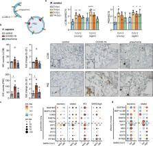
          Viruses manipulate cellular metabolism and macromolecule recycling processes like autophagy. Dysregulated metabolism might lead to excessive inflammatory and autoimmune responses as observed in severe and long COVID-19 patients. Here we show that SARS-CoV-2 modulates cellular metabolism and reduces autophagy. Accordingly, compound-driven induction of autophagy limits SARS-CoV-2 propagation. In detail, SARS-CoV-2-infected cells show accumulation of key metabolites, activation of autophagy inhibitors (AKT1, SKP2) and reduction of proteins responsible for autophagy initiation (AMPK, TSC2, ULK1), membrane nucleation, and phagophore formation (BECN1, VPS34, ATG14), as well as autophagosome-lysosome fusion (BECN1, ATG14 oligomers). Consequently, phagophore-incorporated autophagy markers LC3B-II and P62 accumulate, which we confirm in a hamster model and lung samples of COVID-19 patients. Single-nucleus and single-cell sequencing of patient-derived lung and mucosal samples show differential transcriptional regulation of autophagy and immune genes depending on cell type, disease duration, and SARS-CoV-2 replication levels. Targeting of autophagic pathways by exogenous administration of the polyamines spermidine and spermine, the selective AKT1 inhibitor MK-2206, and the BECN1-stabilizing anthelmintic drug niclosamide inhibit SARS-CoV-2 propagation in vitro with IC 50 values of 136.7, 7.67, 0.11, and 0.13 μM, respectively. Autophagy-inducing compounds reduce SARS-CoV-2 propagation in primary human lung cells and intestinal organoids emphasizing their potential as treatment options against COVID-19.

          Abstract

          Viruses manipulate host cell pathways to support infection. Here the authors show that SARS-CoV-2 infection modulates cellular metabolism and limits autophagy, and identify druggable host pathways for virus inhibition.

          Related collections

          Most cited references83

          • Record: found
          • Abstract: found
          • Article: not found

          Analysis of relative gene expression data using real-time quantitative PCR and the 2(-Delta Delta C(T)) Method.

          The two most commonly used methods to analyze data from real-time, quantitative PCR experiments are absolute quantification and relative quantification. Absolute quantification determines the input copy number, usually by relating the PCR signal to a standard curve. Relative quantification relates the PCR signal of the target transcript in a treatment group to that of another sample such as an untreated control. The 2(-Delta Delta C(T)) method is a convenient way to analyze the relative changes in gene expression from real-time quantitative PCR experiments. The purpose of this report is to present the derivation, assumptions, and applications of the 2(-Delta Delta C(T)) method. In addition, we present the derivation and applications of two variations of the 2(-Delta Delta C(T)) method that may be useful in the analysis of real-time, quantitative PCR data. Copyright 2001 Elsevier Science (USA).
            Bookmark
            • Record: found
            • Abstract: found
            • Article: not found

            Comprehensive Integration of Single-Cell Data

            Single-cell transcriptomics has transformed our ability to characterize cell states, but deep biological understanding requires more than a taxonomic listing of clusters. As new methods arise to measure distinct cellular modalities, a key analytical challenge is to integrate these datasets to better understand cellular identity and function. Here, we develop a strategy to "anchor" diverse datasets together, enabling us to integrate single-cell measurements not only across scRNA-seq technologies, but also across different modalities. After demonstrating improvement over existing methods for integrating scRNA-seq data, we anchor scRNA-seq experiments with scATAC-seq to explore chromatin differences in closely related interneuron subsets and project protein expression measurements onto a bone marrow atlas to characterize lymphocyte populations. Lastly, we harmonize in situ gene expression and scRNA-seq datasets, allowing transcriptome-wide imputation of spatial gene expression patterns. Our work presents a strategy for the assembly of harmonized references and transfer of information across datasets.
              Bookmark
              • Record: found
              • Abstract: found
              • Article: not found

              Integrating single-cell transcriptomic data across different conditions, technologies, and species

              Computational single-cell RNA-seq (scRNA-seq) methods have been successfully applied to experiments representing a single condition, technology, or species to discover and define cellular phenotypes. However, identifying subpopulations of cells that are present across multiple data sets remains challenging. Here, we introduce an analytical strategy for integrating scRNA-seq data sets based on common sources of variation, enabling the identification of shared populations across data sets and downstream comparative analysis. We apply this approach, implemented in our R toolkit Seurat (http://satijalab.org/seurat/), to align scRNA-seq data sets of peripheral blood mononuclear cells under resting and stimulated conditions, hematopoietic progenitors sequenced using two profiling technologies, and pancreatic cell 'atlases' generated from human and mouse islets. In each case, we learn distinct or transitional cell states jointly across data sets, while boosting statistical power through integrated analysis. Our approach facilitates general comparisons of scRNA-seq data sets, potentially deepening our understanding of how distinct cell states respond to perturbation, disease, and evolution.
                Bookmark

                Author and article information

                Contributors
                nils.gassen@ukbonn.de
                marcel.mueller@charite.de
                Journal
                Nat Commun
                Nat Commun
                Nature Communications
                Nature Publishing Group UK (London )
                2041-1723
                21 June 2021
                21 June 2021
                2021
                : 12
                : 3818
                Affiliations
                [1 ]GRID grid.10388.32, ISNI 0000 0001 2240 3300, Department of Psychiatry and Psychotherapy, , University of Bonn, Medical Faculty, ; Bonn, Germany
                [2 ]GRID grid.6363.0, ISNI 0000 0001 2218 4662, Institute of Virology, Charité-Universitätsmedizin Berlin, corporate member of Freie Universität Berlin, Humboldt-Universität zu Berlin, ; Berlin, Germany
                [3 ]GRID grid.452463.2, German Center for Infection Research (DZIF), partner site Charité, ; Berlin, Germany
                [4 ]GRID grid.419502.b, ISNI 0000 0004 0373 6590, Max Planck Institute for Biology of Ageing, ; Cologne, Germany
                [5 ]GRID grid.7468.d, ISNI 0000 0001 2248 7639, Center for Digital Health, , Berlin Institute of Health (BIH) and Charité-Universitätsmedizin Berlin, corporate member of Freie Universität Berlin, Humboldt-Universität zu Berlin, ; Berlin, Germany
                [6 ]GRID grid.14095.39, ISNI 0000 0000 9116 4836, Institute of Virology, Freie Universität Berlin, ; Berlin, Germany
                [7 ]GRID grid.6363.0, ISNI 0000 0001 2218 4662, Molecular Imaging of Immunoregulation, Medizinische Klinik m.S. Infektiologie & Pneumologie, Charité-Universitätsmedizin Berlin, ; Berlin, Germany
                [8 ]GRID grid.6363.0, ISNI 0000 0001 2218 4662, Department of Neuropathology, , Charité-Universitätsmedizin Berlin, corporate member of Freie Universität Berlin and Humboldt-Universität zu Berlin, ; Berlin, Germany
                [9 ]GRID grid.419491.0, ISNI 0000 0001 1014 0849, Berlin Institute for Medical Systems Biology, Max-Delbrück-Center for Molecular Medicine in the Helmholtz Association, ; Berlin, Germany
                [10 ]GRID grid.9764.c, ISNI 0000 0001 2153 9986, Laboratory of Infection Oncology, , Institute of Clinical Molecular Biology, UKSH, Christian Albrechts University of Kiel, ; Kiel, Germany
                [11 ]German Center for Neurodegenerative Diseases (DZNE) Berlin, Berlin, Germany
                [12 ]Cluster of Excellence, NeuroCure, Berlin, Germany
                [13 ]GRID grid.7468.d, ISNI 0000 0001 2248 7639, IRI Life Sciences, Institut für Biologie, Humboldt-Universität zu Berlin, ; Berlin, Germany
                [14 ]GRID grid.6363.0, ISNI 0000 0001 2218 4662, Institute for Pathology, Charité-Universitätsmedizin Berlin, corporate member of Freie Universität Berlin, Humboldt-Universität zu Berlin, ; Berlin, Germany
                [15 ]GRID grid.7497.d, ISNI 0000 0004 0492 0584, German Cancer Consortium (DKTK) Partner Site Berlin, German Cancer Research Center (DKFZ), ; Heidelberg, Germany
                [16 ]GRID grid.35030.35, ISNI 0000 0004 1792 6846, Department of Infectious Diseases and Public Health, Jockey Club College of Veterinary Medicine and Life Sciences, City University of Hong Kong, ; Kowloon Tong, Hong Kong
                [17 ]GRID grid.452624.3, German Center for Lung Research (DZL), ; Berlin, Germany
                [18 ]GRID grid.5253.1, ISNI 0000 0001 0328 4908, Data Science Unit, , Heidelberg University Hospital and BioQuant, ; Heidelberg, Germany
                [19 ]GRID grid.448878.f, ISNI 0000 0001 2288 8774, Martsinovsky Institute of Medical Parasitology, Tropical and Vector Borne Diseases, Sechenov University, ; Moscow, Russia
                Author information
                http://orcid.org/0000-0002-4265-3398
                http://orcid.org/0000-0002-6246-5255
                http://orcid.org/0000-0001-5945-9957
                http://orcid.org/0000-0003-1616-0810
                http://orcid.org/0000-0003-2428-1065
                http://orcid.org/0000-0002-2101-6146
                http://orcid.org/0000-0001-7103-9621
                http://orcid.org/0000-0002-9884-1806
                http://orcid.org/0000-0002-1897-6365
                http://orcid.org/0000-0001-9816-8917
                http://orcid.org/0000-0002-3605-0136
                http://orcid.org/0000-0002-1075-8734
                http://orcid.org/0000-0002-2553-9999
                http://orcid.org/0000-0002-0034-4036
                http://orcid.org/0000-0001-6941-3397
                http://orcid.org/0000-0001-7923-0519
                http://orcid.org/0000-0003-2242-5117
                Article
                24007
                10.1038/s41467-021-24007-w
                8217552
                34155207
                2168a08e-baa3-4b75-ae1c-8ebad912629b
                © The Author(s) 2021

                Open Access This article is licensed under a Creative Commons Attribution 4.0 International License, which permits use, sharing, adaptation, distribution and reproduction in any medium or format, as long as you give appropriate credit to the original author(s) and the source, provide a link to the Creative Commons license, and indicate if changes were made. The images or other third party material in this article are included in the article’s Creative Commons license, unless indicated otherwise in a credit line to the material. If material is not included in the article’s Creative Commons license and your intended use is not permitted by statutory regulation or exceeds the permitted use, you will need to obtain permission directly from the copyright holder. To view a copy of this license, visit http://creativecommons.org/licenses/by/4.0/.

                History
                : 12 March 2021
                : 29 May 2021
                Funding
                Funded by: FundRef https://doi.org/10.13039/501100002347, Bundesministerium für Bildung und Forschung (Federal Ministry of Education and Research);
                Award ID: 01KI20434A
                Award ID: Camo-Covid-19
                Award ID: 01KI1723A
                Award ID: 01KI20434B
                Award Recipient :
                Funded by: COVID-19 funds made available through the Berlin University Alliance and Freie Universität Berlin
                Funded by: Argelander Grant awarded by the University of Bonn
                Funded by: FundRef https://doi.org/10.13039/501100001659, Deutsche Forschungsgemeinschaft (German Research Foundation);
                Award ID: SFB-TR 84, A07
                Award Recipient :
                Categories
                Article
                Custom metadata
                © The Author(s) 2021

                Uncategorized
                macroautophagy,sars-cov-2
                Uncategorized
                macroautophagy, sars-cov-2

                Comments

                Comment on this article