22
views
0
recommends
+1 Recommend
0 collections
    0
    shares
      • Record: found
      • Abstract: found
      • Article: found
      Is Open Access

      Immunomodulatory Activity of Recombinant Ricin Toxin Binding Subunit B (RTB)

      research-article

      Read this article at

      Bookmark
          There is no author summary for this article yet. Authors can add summaries to their articles on ScienceOpen to make them more accessible to a non-specialist audience.

          Abstract

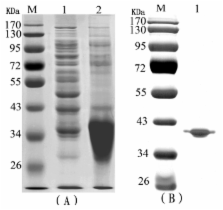
          Ricin toxin binding subunit B (RTB) is one of the subunits of the ricin protein. RTB has been used as adjuvant, but little is known about its mechanism. In this study, we found that RTB increased not only nitric oxide (NO) release, but also tumor necrosis factor (TNF)-α and interleukin (IL)-6 production in mouse macrophage cell line RAW264.7 cells. They subsequently exhibited enhanced ConA-induced T-cell and LPS-induced B-cell proliferative responses. We also examined the cytokines that were produced from splenocytes following in vitro RTB administration. Increased levels of IL-2, interferon (IFN)-γ and TNF-α were observed, while IL-4 and IL-5 were unaffected. These results demonstrate that recombinant RTB can act on the immune system and activate T-cells by introducing a Th1 immune response. Th1 cells might be the primary cellular target affected by RTB. Our results suggest that the recombinant RTB can promote the activation of macrophages and has a beneficial effect on immunomodulatory activity.

          Related collections

          Most cited references23

          • Record: found
          • Abstract: found
          • Article: not found

          Two types of murine helper T cell clone. I. Definition according to profiles of lymphokine activities and secreted proteins.

          A panel of antigen-specific mouse helper T cell clones was characterized according to patterns of lymphokine activity production, and two types of T cell were distinguished. Type 1 T helper cells (TH1) produced IL 2, interferon-gamma, GM-CSF, and IL 3 in response to antigen + presenting cells or to Con A, whereas type 2 helper T cells (TH2) produced IL 3, BSF1, and two other activities unique to the TH2 subset, a mast cell growth factor distinct from IL 3 and a T cell growth factor distinct from IL 2. Clones representing each type of T cell were characterized, and the pattern of lymphokine activities was consistent within each set. The secreted proteins induced by Con A were analyzed by biosynthetic labeling and SDS gel electrophoresis, and significant differences were seen between the two groups of T cell line. Both types of T cell grew in response to alternating cycles of antigen stimulation, followed by growth in IL 2-containing medium. Examples of both types of T cell were also specific for or restricted by the I region of the MHC, and the surface marker phenotype of the majority of both types was Ly-1+, Lyt-2-, L3T4+, Both types of helper T cell could provide help for B cells, but the nature of the help differed. TH1 cells were found among examples of T cell clones specific for chicken RBC and mouse alloantigens. TH2 cells were found among clones specific for mouse alloantigens, fowl gamma-globulin, and KLH. The relationship between these two types of T cells and previously described subsets of T helper cells is discussed.
            Bookmark
            • Record: found
            • Abstract: found
            • Article: not found

            A potential role of macrophage activation in the treatment of cancer.

            One of the functions of macrophages is to provide a defense mechanism against tumor cells. In the last decades the mechanism of tumor cell killing by macrophages have been studied extensively. The tumor cytotoxic function of macrophages requires stimulation either with bacterial cell wall products such as lipopolysaccharide (LPS) or muramyldipeptide (MDP) or with cytokines such as interferon-gamma (IFN-gamma) and granulocyte-macrophage colony-stimulating factor (GM-CSF). Activated macrophages secrete several substances that are directly involved in tumor cell killing i.e. tumor necrosis factor (TNF) and nitric oxide (NO). On the other hand, substances are secreted that are able to stimulate tumor cell growth, depending on the stage and the nature of the tumor. Several clinical trials have been performed aiming at the activation of macrophages or dendritic cells, a subpopulation of the macrophages. In this review we will summarize and discuss experimental studies and clinical trials based on the activation of macrophages.
              Bookmark
              • Record: found
              • Abstract: found
              • Article: not found

              The history of ricin, abrin and related toxins.

              Ricin, abrin and related plant toxins have played interesting and important roles in the history of clinical medicine and biomedical research. The use of these proteins in medical treatment since ancient times is reviewed. Later the proteins played important roles in the early days of immunological research and some of the fundamental principles of immunology were discovered with toxic proteins of this group. During the last three decades the mechanism of action of the toxins was elucidated. This led to a major effort to target the toxins to malignant cells. Ricin has been used in bioterrorism. Recently, the toxins have played important roles as experimental models to elucidate the intracellular trafficking of endocytosed proteins.
                Bookmark

                Author and article information

                Journal
                Int J Mol Sci
                Int J Mol Sci
                ijms
                International Journal of Molecular Sciences
                Molecular Diversity Preservation International (MDPI)
                1422-0067
                June 2013
                13 June 2013
                : 14
                : 6
                : 12401-12410
                Affiliations
                [1 ]Institute of Military Veterinary, Academy of Military Medical Sciences, Zoonosis Prevention and Control Key Laboratory, Changchun 130122, China; E-Mails: xuna206@ 123456aliyun.com (N.X.); lisongyan888@ 123456163.com (S.L.); liulinna@ 123456126.com (L.L.); puzhaoyang@ 123456126.com (Z.P.); jywan_1973@ 123456163.com (J.W.)
                [2 ]Department of Immunology, Norman Bethune College of Medical Science, Jilin University, Changchun 130021, China; E-Mails: yuanhongyan@ 123456126.com (H.Y.); wanghuiwen@ 123456126.com (H.W.)
                [3 ]Dean’s Office, Jilin Medical College, Jilin 132013, China
                [4 ]Animal Laboratory Center, 302 Hospital of People’s Liberation Army, Beijing 100039, China
                Author notes
                [†]

                These authors contributed equally to this work.

                [* ]Authors to whom correspondence should be addressed; E-Mails: liuws85952@ 123456163.com (W.L.); changyp@ 123456jlu.edu.cn (Y.C.); lrsheng@ 123456sohu.com (R.L.); Tel./Fax: +86-431-8698-5951 (W.L.); +86-431-8561-9476 (Y.C.); +86-10-6693-3326 (R.L.).
                Article
                ijms-14-12401
                10.3390/ijms140612401
                3709792
                23765218
                2211453c-29ad-41d6-851c-a5eb8fdc49a7
                © 2013 by the authors; licensee MDPI, Basel, Switzerland

                This article is an open access article distributed under the terms and conditions of the Creative Commons Attribution license ( http://creativecommons.org/licenses/by/3.0/).

                History
                : 04 February 2013
                : 07 May 2013
                : 24 May 2013
                Categories
                Article

                Molecular biology
                rtb,macrophage,lymphocyte proliferation,th1 cell,cytokine
                Molecular biology
                rtb, macrophage, lymphocyte proliferation, th1 cell, cytokine

                Comments

                Comment on this article