28
views
0
recommends
+1 Recommend
0 collections
    0
    shares
      • Record: found
      • Abstract: found
      • Article: not found

      Small field-of-view dedicated cardiac SPECT systems: impact of projection truncation

      research-article

      Read this article at

      ScienceOpenPublisherPMC
      Bookmark
          There is no author summary for this article yet. Authors can add summaries to their articles on ScienceOpen to make them more accessible to a non-specialist audience.

          Abstract

          Purpose

          Small field-of-view (FOV) dedicated cardiac SPECT systems suffer from truncated projection data. This results in (1) neglect of liver activity that otherwise could be used to estimate (and subsequently correct) the amount of scatter in the myocardium by model-based scatter correction, and (2) distorted attenuation maps. In this study, we investigated to what extent truncation impacts attenuation correction and model-based scatter correction in the cases of 99mTc, 201Tl, and simultaneous 99mTc/ 201Tl studies. In addition, we evaluated a simple correction method to mitigate the effects of truncation.

          Methods

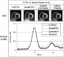
          Digital thorax phantoms of different sizes were used to simulate the full FOV SPECT projections for 99mTc, 201Tl, and simultaneous 99mTc/ 201Tl studies. Small FOV projections were obtained by artificially truncating the full FOV projections. Deviations from ideal heart positioning were simulated by axially shifting projections resulting in more severe liver truncation. Effects of truncation on SPECT images were tested for ordered subset (OS) expectation maximization reconstruction with (1) attenuation correction and detector response modelling (OS-AD), and (2) with additional Monte-Carlo-based scatter correction (OS-ADS). To correct truncation-induced artefacts, we axially extended truncated projections on both sides by duplicating pixel values on the projection edge.

          Results

          For both 99mTc and 201Tl, differences in the reconstructed myocardium between full FOV and small FOV projections were negligible. In the nine myocardial segments, the maximum deviations of the average pixel values were 1.3% for OS-AD and 3.5% for OS-ADS. For the simultaneous 99mTc/ 201Tl studies, reconstructed 201Tl SPECT images from full FOV and small FOV projections showed clearly different image profiles due to truncation. The maximum deviation in defected segments was found to be 49% in the worst-case scenario. However, artificially extending projections reduced deviations in defected segments to a few percent.

          Conclusion

          Our results indicate that, for single isotope studies, using small FOV systems has little impact on attenuation correction and model-based scatter correction. For simultaneous 99mTc/ 201Tl studies, artificial projection extension almost fully eliminates the adverse effects of projection truncation.

          Related collections

          Most cited references34

          • Record: found
          • Abstract: found
          • Article: not found

          Efficient fully 3-D iterative SPECT reconstruction with Monte Carlo-based scatter compensation.

          Quantitative accuracy of single photon emission computed tomography (SPECT) images is highly dependent on the photon scatter model used for image reconstruction. Monte Carlo simulation (MCS) is the most general method for detailed modeling of scatter, but to date, fully three-dimensional (3-D) MCS-based statistical SPECT reconstruction approaches have not been realized, due to prohibitively long computation times and excessive computer memory requirements. MCS-based reconstruction has previously been restricted to two-dimensional approaches that are vastly inferior to fully 3-D reconstruction. Instead of MCS, scatter calculations based on simplified but less accurate models are sometimes incorporated in fully 3-D SPECT reconstruction algorithms. We developed a computationally efficient fully 3-D MCS-based reconstruction architecture by combining the following methods: 1) a dual matrix ordered subset (DM-OS) reconstruction algorithm to accelerate the reconstruction and avoid massive transition matrix precalculation and storage; 2) a stochastic photon transport calculation in MCS is combined with an analytic detector modeling step to reduce noise in the Monte Carlo (MC)-based reprojection after only a small number of photon histories have been tracked; and 3) the number of photon histories simulated is reduced by an order of magnitude in early iterations, or photon histories calculated in an early iteration are reused. For a 64 x 64 x 64 image array, the reconstruction time required for ten DM-OS iterations is approximately 30 min on a dual processor (AMD 1.4 GHz) PC, in which case the stochastic nature of MCS modeling is found to have a negligible effect on noise in reconstructions. Since MCS can calculate photon transport for any clinically used photon energy and patient attenuation distribution, the proposed methodology is expected to be useful for obtaining highly accurate quantitative SPECT images within clinically acceptable computation times.
            Bookmark
            • Record: found
            • Abstract: found
            • Article: not found

            A new algorithm for the quantitation of myocardial perfusion SPECT. I: technical principles and reproducibility.

            We have developed a new, completely automatic 3-dimensional software approach to quantitative perfusion SPECT. The main features of the software are myocardial sampling based on an ellipsoid model; use of the entire count profile between the endocardial and epicardial surfaces; independence of the algorithm from myocardial shape, size, and orientation and establishment of a standard 3-dimensional point-to-point correspondence among all sampled myocardial regions; automatic generation of quantitative measurements and 5-point semiquantitative scores for each of 20 myocardial segments and automatic derivation of summed perfusion scores; and automatic generation of normal limits for any given patient population on the basis of data fractionally normalized to minimize hot spot artifacts. The new algorithm was tested on the tomographic images of 420 patients studied with a rest 201TI (111-167 MBq, 35 s/projection)-stress 99mTc-sestamibi (925-1480 MBq, 25 s/projection) separate dual-isotope protocol on a single-detector camera, a dual-detector 90 degrees camera, and a triple-detector camera. The algorithm was successful in 397 of 420 patients (94.5%) and 816 of 840 image datasets (97.1%), with a statistically significant difference between the success rates of the 201TI images (399/ 420, or 95.0%) and the 99mTc images (417/420, or 99.3%; P < 0.001). Algorithm failure was caused by extracardiac uptake (10/24, or 41.7%) or inaccurate identification of the valve plane because of low count statistics (14/24, or 58.3%) and was obviated by simply limiting the image volume in which the software operates. Reproducibility of measurements of summed perfusion scores (r = 0.999 and 1 for stress and rest, respectively), global defect extent (r = 0.999 and 1 for stress and rest, respectively), and segmental perfusion scores (exact agreement = 99.9%, kappa = 0.998 for stress and 0.997 for rest) was extremely high. Automatic 3-dimensional quantitation of perfusion from 201Tl and 99mTc-sestamibi images is feasible and reproducible. The described software, because it is based on the same sampling scheme used for gated SPECT analysis, ensures intrinsically perfect registration of quantitative perfusion with quantitative regional wall motion and thickening information, if gated SPECT is used.
              Bookmark
              • Record: found
              • Abstract: found
              • Article: not found

              Accelerated iterative transmission CT reconstruction using an ordered subsets convex algorithm.

              Iterative maximum likelihood (ML) transmission computed tomography algorithms have distinct advantages over Fourier-based reconstruction, but unfortunately require increased computation time. The convex algorithm [1] is a relatively fast iterative ML algorithm but it is nevertheless too slow for many applications. Therefore, an acceleration of this algorithm by using ordered subsets of projections is proposed [ordered subsets convex algorithm (OSC)]. OSC applies the convex algorithm sequentially to subsets of projections. OSC was compared with the convex algorithm using simulated and physical thorax phantom data. Reconstructions were performed for OSC using eight and 16 subsets (eight and four projections/subset, respectively). Global errors, image noise, contrast recovery, and likelihood increase were calculated. Results show that OSC is faster than the convex algorithm, the amount of acceleration being approximately proportional to the number of subsets in OSC, and it causes only a slight increase of noise and global errors in the reconstructions. Images and image profiles of the reconstructions were in good agreement. In conclusion, OSC and the convex algorithm result in similar image quality but OSC is more than an order of magnitude faster.
                Bookmark

                Author and article information

                Contributors
                +31-30-2538846 , +31-30-2539032 , j.xiao@robeco.nl
                Journal
                Eur J Nucl Med Mol Imaging
                European Journal of Nuclear Medicine and Molecular Imaging
                Springer-Verlag (Berlin/Heidelberg )
                1619-7070
                1619-7089
                1 September 2009
                1 September 2009
                March 2010
                : 37
                : 3
                : 528-536
                Affiliations
                [1 ]Image Sciences Institute, University Medical Centre Utrecht, Universiteitsweg 100, STR 5.203, 3584 CG Utrecht, The Netherlands
                [2 ]Robeco, Coolsingel 120, Rotterdam, 3011 AG The Netherlands
                [3 ]Department of Nuclear Medicine, St. Antonius Hospital, P.O. Box 2500, 3430 EM Nieuwegein, The Netherlands
                [4 ]Section of Radiation, Detection and Medical Imaging, Delft University of Technology, Mekelweg 15, Delft, The Netherlands
                Article
                1223
                10.1007/s00259-009-1223-9
                2822234
                19722106
                22b77092-99a7-4e2a-af85-562f79fdac91
                © The Author(s) 2009
                History
                : 24 February 2009
                : 1 July 2009
                Categories
                Original Article
                Custom metadata
                © Springer-Verlag 2010

                Radiology & Imaging
                small field-of-view,scatter correction,cardiac spect,truncation,attenuation correction

                Comments

                Comment on this article