7
views
0
recommends
+1 Recommend
0 collections
    0
    shares
      • Record: found
      • Abstract: found
      • Article: found
      Is Open Access

      Fabrication of a low-cost strap for holding precordial electrodes on the hirsute chest

      research-article

      Read this article at

      Bookmark
          There is no author summary for this article yet. Authors can add summaries to their articles on ScienceOpen to make them more accessible to a non-specialist audience.

          Abstract

          Background:

          Reusable suction-cup electrodes are used for recording a 12-lead electrocardiogram (ECG) in resource-limited settings. These electrodes may easily detach if those are attached on a hirsute chest. Additionally, the suction pressure may cause erythema and pain.

          Aim:

          The aim of this study was to develop a low-cost strap for holding the suction-cup-based precordial electrodes and to test its applicability to the recording of ECG.

          Materials and Methods:

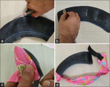
          A scrap rubber tube was cut in size so that it can cover all the precordial electrode positions. Slit openings (electrodes can be inserted through these opening) were made on this rubber strap. A cloth and a hook-and-loop fastener were used to make an adjustable fastener. ECG was recorded first on 16 non-hairy males with electrodes placed on the chest with the strap and then with electrodes attached by suction. After that, ECG was recorded on 16 males with hirsute chest first with the electrodes placed with the help of the strap and then with suction (without strap) on the shaved chest.

          Results:

          The outcome of this study is a low-cost ECG chest strap for a hirsute chest. Both the negative and positive QRS voltages in six precordial leads recorded with electrodes placed with the strap were similar to that of suction-attached electrodes in both non-hairy and hirsute chest.

          Conclusion:

          Developed rubber chest strap can be made with minimal cost and expertise in any resource-limited settings. This would help in recording the ECG of a patient with a hirsute chest. This prototype strap has scope for further improvement.

          Related collections

          Most cited references10

          • Record: found
          • Abstract: found
          • Article: not found

          High and dry? Comparing active dry EEG electrodes to active and passive wet electrodes.

          Dry electrodes are becoming popular for both lab-based and consumer-level electrophysiological-recording technologies because they better afford the ability to move traditional lab-based research into the real world. It is unclear, however, how dry electrodes compare in data quality to traditional electrodes. The current study compared three EEG electrode types: (a) passive-wet electrodes with no onboard amplification, (b) actively amplified, wet electrodes with moderate impedance levels, and low impedance levels, and (c) active-dry electrodes with very high impedance. Participants completed a classic P3 auditory oddball task to elicit characteristic EEG signatures and event-related potentials (ERPs). Across the three electrode types, we compared single-trial noise, average ERPs, scalp topographies, ERP noise, and ERP statistical power as a function of number of trials. We extended past work showing active electrodes' insensitivity to moderate levels of interelectrode impedance when compared to passive electrodes in the same amplifier. Importantly, the new dry electrode system could reliably measure EEG spectra and ERP components comparable to traditional electrode types. As expected, however, dry active electrodes with very high interelectrode impedance exhibited marked increases in single-trial and average noise levels, which decreased statistical power, requiring more trials to detect significant effects. This power decrease must be considered as a trade-off with the ease of application and long-term use. The current results help set constraints on experimental design with novel dry electrodes, and provide important evidence needed to measure brain activity in novel settings and situations.
            Bookmark
            • Record: found
            • Abstract: found
            • Article: not found

            Electrocardiography in primary care; is it useful?

            Little is known about the value of electrocardiography in primary care. To assess whether electrocardiography (ECG) is a useful instrument, in addition to history taking and physical examination, in that it changes the general practitioner's management of patients with suspected cardiovascular symptoms or disease. We performed a prospective study in a group practice of eight general practitioners in The Netherlands. During 2 years all ECGs that were recorded in these practices were studied. Two questionnaires were filled out by the general practitioners, one before and one after the ECG recording, to determine indication for electrocardiography, the general practitioner's anticipated management before and after ECG results, and the subjective usefulness according to the applying doctor. All ECGs were reviewed by an experienced general practitioner working in the group practice and later on by a cardiologist. In addition, all clinical information, including the 6 months follow-up period, was scrutinised by both the cardiologist and general practitioner to establish the patients' prognosis. A total of 301 ECGs was included in the analysis. Main indications for electrocardiography were chest pain (57%), and collapse or palpitation (30%). In 92 (30.6%; 95% CI 25.4-35.8) patients a change in management by the general practitioner occurred following the ECG results. Most prevalent changes were non-referral to a cardiologist, while referral was anticipated before the ECG results (34%), referral while the patient would not have been referred without ECG results (20%), and change in cardiovascular therapy (40%). In one of these cases only, this change could be considered unfavourable, since a subendocardial infarction, not detectable on the ECG, was missed. In patients with chest pain, a normal ECG (likelihood ratio (LR) 0.06) and an abnormal ECG (LR 13.3) were very useful to distinguish between patients likely or unlikely to experience cardiac events in the near future. The mean subjective usefulness, on a scale of 0-100, of the ECG evaluation according to the applying general practitioner was 77. 5 (S.D. 14.4). There was good agreement in ECG interpretation between the experienced general practitioner, the cardiologist and a second general practitioner. Electrocardiography in addition to history taking and physical examination, may be an important tool in primary care. It can reduce considerably the number of unnecessary referrals.
              Bookmark
              • Record: found
              • Abstract: found
              • Article: found
              Is Open Access

              Microbial contamination of manually reprocessed, ready to use ECG lead wire in intensive care units

              Background: A number of studies have shown that non-critical medical devices can be contaminated with pathogens, including those resistant to antibiotics and thus become a potential vector for transmission. Electrocardiography (ECG) lead wire are non-critical medical device which are always attached on patient skin during their stay in intensive care unit (ICU). In view of the patient’s critical conditions and exposure to invasive procedures, identification and prevention of possible risks are important to prevent infection in ICUs. Objective: The objective of this study was to determine the presence of bacterial and fungal contamination on cleaned and disinfected reusable ECG lead wires in intensive care units in a hospital. Methods: A total of 408 cleaned ECG lead wires from 93 bed-side ECG devices and 43 ECG lead wires from 5 portable ECG devices from 4 intensive care units (ICUs) and 1 post-anaesthesia care unit (PACU) were sampled. ECG lead wires were stirred in 0.89% NaCl with added neutralizer for 30 seconds. Samples of the solutions were cultured directly on blood agar. The remaining solution was cultured on blood agar after sterile filtration. The number of colony forming units (CFUs) was counted and the microorganisms were identified. Results: More than half of examined ECG lead wires (n=232; 51.4%) were contaminated with >30 CFUs/mL sample of bacteria or with risk pathogens. Gram-positive bacteria were the most frequently isolated organisms; particularly, coagulase negative staphylococci (96%) and aerobic spore forming bacteria (71.2%). Compared to ICUs, PACU had significantly lower proportion of contaminated ECG lead wires (p<0.05). The proportion of contaminated ECG lead wires, as well as mean number of cfus per ECG lead wire, was also significantly lower among multi-wire ECG leads compared to single-wire ECG leads. Conclusions: Manually cleaned ECG lead wires may serve as a vector for transmission of nosocomial pathogens. The current reprocessing technique for ECG lead wires needs to be improved.
                Bookmark

                Author and article information

                Journal
                J Family Med Prim Care
                J Family Med Prim Care
                JFMPC
                Journal of Family Medicine and Primary Care
                Wolters Kluwer - Medknow (India )
                2249-4863
                2278-7135
                May 2020
                31 May 2020
                : 9
                : 5
                : 2359-2363
                Affiliations
                [1 ] Department of Physiology, Bhima Bhoi Medical College and Hospital, Balangir, Odisha, India
                [2 ] Department of Medicine, Ramakrishna Mission Seva Pratishthan, Vivekananda Institute of Medical Sciences, West Bengal, India
                [3 ] Department of Physiology, Raiganj Government Medical College and Hospital, West Bengal, India
                [4 ] Department of Physiology, Fakir Mohan Medical College and Hospital, Balasore, Odisha, India
                [5 ] Department of Biochemistry, Fakir Mohan Medical College and Hospital, Balasore, Odisha, India
                Author notes
                Address for correspondence: Dr. Tanmoy Chattopadhyay, Department of Medicine, Vivekananda Institute of Medical Sciences, 99 Sarat Bose Road, West Bengal - 700 026, India. E-mail: himelmkcg@ 123456gmail.com
                Article
                JFMPC-9-2359
                10.4103/jfmpc.jfmpc_292_20
                7380826
                32754501
                22f0317b-2c5b-4baf-ac4b-7646cd3f4dcc
                Copyright: © 2020 Journal of Family Medicine and Primary Care

                This is an open access journal, and articles are distributed under the terms of the Creative Commons Attribution-NonCommercial-ShareAlike 4.0 License, which allows others to remix, tweak, and build upon the work non-commercially, as long as appropriate credit is given and the new creations are licensed under the identical terms.

                History
                : 21 February 2020
                : 14 March 2020
                : 26 March 2020
                Categories
                Original Article

                chest lead,ecg strap,ekg,electrocardiography,electrodes,erythema,innovation

                Comments

                Comment on this article