32
views
0
recommends
+1 Recommend
0 collections
    0
    shares
      • Record: found
      • Abstract: found
      • Article: found
      Is Open Access

      Microbial recognition by GEF-H1 controls IKKε mediated activation of IRF5

      research-article

      Read this article at

      Bookmark
          There is no author summary for this article yet. Authors can add summaries to their articles on ScienceOpen to make them more accessible to a non-specialist audience.

          Abstract

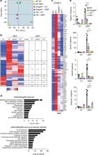
          During infection, transcription factor interferon regulatory factor 5 (IRF5) is essential for the control of host defense. Here we show that the microtubule-associated guanine nucleotide exchange factor (GEF)-H1, is required for the phosphorylation of IRF5 by microbial muramyl-dipeptides (MDP), the minimal structural motif of peptidoglycan of both Gram-positive and Gram-negative bacteria. Specifically, GEF-H1 functions in a microtubule based recognition system for microbial peptidoglycans that mediates the activation of IKKε which we identify as a new upstream IKKα/β and IRF5 kinase. The deletion of GEF-H1 or dominant-negative variants of GEF-H1 prevent activation of IKKε and phosphorylation of IRF5. The GEF-H1-IKKε-IRF5 signaling axis functions independent of NOD-like receptors and is critically required for the recognition of intracellular peptidoglycans and host defenses against Listeria monocytogenes.

          Abstract

          The transcription factor IRF5 is essential for immune defense against pathogens. Here, the authors show that the microtubule-associated factor GEF-H1 plays a critical role in host defense against Listeria monocytogenes in macrophages via activation of the IRF5 kinase IKKε.

          Related collections

          Most cited references38

          • Record: found
          • Abstract: found
          • Article: not found

          IRF5 promotes inflammatory macrophage polarization and TH1-TH17 responses.

          Polymorphisms in the gene encoding the transcription factor IRF5 that lead to higher mRNA expression are associated with many autoimmune diseases. Here we show that IRF5 expression in macrophages was reversibly induced by inflammatory stimuli and contributed to the plasticity of macrophage polarization. High expression of IRF5 was characteristic of M1 macrophages, in which it directly activated transcription of the genes encoding interleukin 12 subunit p40 (IL-12p40), IL-12p35 and IL-23p19 and repressed the gene encoding IL-10. Consequently, those macrophages set up the environment for a potent T helper type 1 (T(H)1)-T(H)17 response. Global gene expression analysis demonstrated that exogenous IRF5 upregulated or downregulated expression of established phenotypic markers of M1 or M2 macrophages, respectively. Our data suggest a critical role for IRF5 in M1 macrophage polarization and define a previously unknown function for IRF5 as a transcriptional repressor.
            Bookmark
            • Record: found
            • Abstract: not found
            • Article: not found

            The IκB Kinase Complex (IKK) Contains Two Kinase Subunits, IKKα and IKKβ, Necessary for IκB Phosphorylation and NF-κB Activation

              Bookmark
              • Record: found
              • Abstract: found
              • Article: not found

              Host defense, viruses and apoptosis.

              To thwart viral infection, the host has developed a formidable and integrated defense network that comprises our innate and adaptive immune response. In recent years, it has become clear that in an attempt to prevent viral replication, viral dissemination or persistent viral infection of the cell, many of these protective measures actually involve the induction of programmed cell death, or apoptosis. An initial response to viral infection primarily involves the innate arm of immunity and the killing of infected cells with cytotoxic lymphocytes such as natural killer (NK) cells through mechanisms that include the employment of perforin and granzymes. Once the virus has invaded the cell, however, a second host defense-mediated response is also triggered which involves the induction of a family of cytokines known as the interferons (IFNs). The IFNs, which are essential for initiating and coordinating a successful antiviral response, function by stimulating the adaptive arm of immunity involving cytotoxic T cells (CTLs), and by inducing a number of intracellular genes that directly prevent virus replication/cytolysis or that facilitate apoptosis. The IFN-induced gene family is now known to comprise the death ligand TRAIL, the dsRNA-dependent protein kinase (PKR), interferon regulatory factors (IRFs) and the promyelocytic leukemia gene (PML), all of which have been reported to be mediators of cell death. That DNA array analyses indicate that numerous cellular genes, many as yet uncharacterized, may similarly be induced by IFN, further emphasizes the likely importance that these cytokines have in the modulation of apoptosis. This likelihood is additionally underlined by the elaborate strategies developed by viruses to inhibit IFN-antiviral function and the mechanisms of cell death.
                Bookmark

                Author and article information

                Contributors
                hans-christian_reinecker@hms.harvard.edu
                Journal
                Nat Commun
                Nat Commun
                Nature Communications
                Nature Publishing Group UK (London )
                2041-1723
                22 March 2019
                22 March 2019
                2019
                : 10
                : 1349
                Affiliations
                ISNI 0000 0004 0386 9924, GRID grid.32224.35, Department of Medicine, Gastrointestinal Unit and Center for the Study of Inflammatory Bowel Disease, , Massachusetts General Hospital, Harvard Medical School, ; Boston, MA 02114 USA
                Author information
                http://orcid.org/0000-0002-7505-1617
                Article
                9283
                10.1038/s41467-019-09283-x
                6430831
                22fda1fc-48ef-4cd4-a9c5-f0473b16d07c
                © The Author(s) 2019

                Open Access This article is licensed under a Creative Commons Attribution 4.0 International License, which permits use, sharing, adaptation, distribution and reproduction in any medium or format, as long as you give appropriate credit to the original author(s) and the source, provide a link to the Creative Commons license, and indicate if changes were made. The images or other third party material in this article are included in the article’s Creative Commons license, unless indicated otherwise in a credit line to the material. If material is not included in the article’s Creative Commons license and your intended use is not permitted by statutory regulation or exceeds the permitted use, you will need to obtain permission directly from the copyright holder. To view a copy of this license, visit http://creativecommons.org/licenses/by/4.0/.

                History
                : 17 July 2018
                : 27 February 2019
                Categories
                Article
                Custom metadata
                © The Author(s) 2019

                Uncategorized
                Uncategorized

                Comments

                Comment on this article