12
views
0
recommends
+1 Recommend
0 collections
    0
    shares
      • Record: found
      • Abstract: found
      • Article: found
      Is Open Access

      Histone demethylase JMJD1A coordinates acute and chronic adaptation to cold stress via thermogenic phospho-switch

      research-article

      Read this article at

      Bookmark
          There is no author summary for this article yet. Authors can add summaries to their articles on ScienceOpen to make them more accessible to a non-specialist audience.

          Abstract

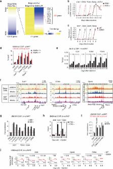
          In acute cold stress in mammals, JMJD1A, a histone H3 lysine 9 (H3K9) demethylase, upregulates thermogenic gene expressions through β-adrenergic signaling in brown adipose tissue (BAT). Aside BAT-driven thermogenesis, mammals have another mechanism to cope with long-term cold stress by inducing the browning of the subcutaneous white adipose tissue (scWAT). Here, we show that this occurs through a two-step process that requires both β-adrenergic-dependent phosphorylation of S265 and demethylation of H3K9me2 by JMJD1A. The histone demethylation-independent acute Ucp1 induction in BAT and demethylation-dependent chronic Ucp1 expression in beige scWAT provides complementary molecular mechanisms to ensure an ordered transition between acute and chronic adaptation to cold stress. JMJD1A mediates two major signaling pathways, namely, β-adrenergic receptor and peroxisome proliferator-activated receptor-γ (PPARγ) activation, via PRDM16-PPARγ-P-JMJD1A complex for beige adipogenesis. S265 phosphorylation of JMJD1A, and the following demethylation of H3K9me2 might prove to be a novel molecular target for the treatment of metabolic disorders, via promoting beige adipogenesis.

          Abstract

          JMJD1A is essential for thermogenic gene induction in brown adipose tissue. Here the authors show that white adipose tissue beige-ing requires both β-adrenergic-dependent phosphorylation of S265 and demethylation activity of JMJD1A while brown adipose tissue-driven thermogenesis requires β-adrenergic dependent phosphorylation of S265 but is independent of H3K9me2 demethylation.

          Related collections

          Most cited references21

          • Record: found
          • Abstract: found
          • Article: not found

          Brown remodeling of white adipose tissue by SirT1-dependent deacetylation of Pparγ.

          Brown adipose tissue (BAT) can disperse stored energy as heat. Promoting BAT-like features in white adipose (WAT) is an attractive, if elusive, therapeutic approach to staunch the current obesity epidemic. Here we report that gain of function of the NAD-dependent deacetylase SirT1 or loss of function of its endogenous inhibitor Deleted in breast cancer-1 (Dbc1) promote "browning" of WAT by deacetylating peroxisome proliferator-activated receptor (Ppar)-γ on Lys268 and Lys293. SirT1-dependent deacetylation of Lys268 and Lys293 is required to recruit the BAT program coactivator Prdm16 to Pparγ, leading to selective induction of BAT genes and repression of visceral WAT genes associated with insulin resistance. An acetylation-defective Pparγ mutant induces a brown phenotype in white adipocytes, whereas an acetylated mimetic fails to induce "brown" genes but retains the ability to activate "white" genes. We propose that SirT1-dependent Pparγ deacetylation is a form of selective Pparγ modulation of potential therapeutic import. Copyright © 2012 Elsevier Inc. All rights reserved.
            Bookmark
            • Record: found
            • Abstract: found
            • Article: not found

            PPARγ agonists induce a white-to-brown fat conversion through stabilization of PRDM16 protein.

            Brown adipose tissue dissipates energy through heat and functions as a defense against cold and obesity. PPARγ ligands have been shown to induce the browning of white adipocytes; however, the underlying mechanisms remain unclear. Here, we show that PPARγ ligands require full agonism to induce a brown fat gene program preferentially in subcutaneous white adipose. These effects require expression of PRDM16, a factor that controls the development of classical brown fat. Depletion of PRDM16 blunts the effects of the PPARγ agonist rosiglitazone on the induced brown fat gene program. Conversely, PRDM16 and rosiglitazone synergistically activate the brown fat gene program in vivo. This synergy is tightly associated with an increased accumulation of PRDM16 protein, due in large measure to an increase in the half-life of the protein in agonist treated cells. Identifying compounds that stabilize PRDM16 protein may represent a plausible therapeutic pathway for the treatment of obesity and diabetes. Copyright © 2012 Elsevier Inc. All rights reserved.
              Bookmark
              • Record: found
              • Abstract: found
              • Article: not found

              Nonshivering thermogenesis and its adequate measurement in metabolic studies.

              Alterations in nonshivering thermogenesis are presently discussed as being both potentially causative of and able to counteract obesity. However, the necessity for mammals to defend their body temperature means that the ambient temperature profoundly affects the outcome and interpretation of metabolic experiments. An adequate understanding and assessment of nonshivering thermogenesis is therefore paramount for metabolic studies. Classical nonshivering thermogenesis is facultative, i.e. it is only activated when an animal acutely requires extra heat (switched on in minutes), and adaptive, i.e. it takes weeks for an increase in capacity to develop. Nonshivering thermogenesis is fully due to brown adipose tissue activity; adaptation corresponds to the recruitment of this tissue. Diet-induced thermogenesis is probably also facultative and adaptive and due to brown adipose tissue activity. Although all mammals respond to injected/infused norepinephrine (noradrenaline) with an increase in metabolism, in non-adapted mammals this increase mainly represents the response of organs not involved in nonshivering thermogenesis; only the increase after adaptation represents nonshivering thermogenesis. Thermogenesis (metabolism) should be expressed per animal, and not per body mass [not even to any power (0.75 or 0.66)]. A 'cold tolerance test' does not examine nonshivering thermogenesis capacity; rather it tests shivering capacity and endurance. For mice, normal animal house temperatures are markedly below thermoneutrality, and the mice therefore have a metabolic rate and food consumption about 1.5 times higher than their intrinsic requirements. Housing and examining mice at normal house temperatures carries a high risk of identifying false positives for intrinsic metabolic changes; in particular, mutations/treatments that affect the animal's insulation (fur, skin) may lead to such problems. Correspondingly, true alterations in intrinsic metabolic rate remain undetected when metabolism is examined at temperatures below thermoneutrality. Thus, experiments with animals kept and examined at thermoneutrality are likely to yield an improved possibility of identifying agents and genes important for human energy balance.
                Bookmark

                Author and article information

                Contributors
                inagaki@lsbm.org
                jmsakai-tky@umin.ac.jp
                Journal
                Nat Commun
                Nat Commun
                Nature Communications
                Nature Publishing Group UK (London )
                2041-1723
                19 April 2018
                19 April 2018
                2018
                : 9
                : 1566
                Affiliations
                [1 ]ISNI 0000 0001 2151 536X, GRID grid.26999.3d, Division of Metabolic Medicine, Research Center for Advanced Science and Technology, , The University of Tokyo, ; Tokyo, 153-8904 Japan
                [2 ]Niigata College of Medical Technology, Niigata, 950-2076 Japan
                [3 ]Rhelixa Inc., Tokyo, 101-0032 Japan
                [4 ]ISNI 0000 0001 2151 536X, GRID grid.26999.3d, Genome Science Division, Research Center for Advanced Science and Technology, , The University of Tokyo, ; Tokyo, 153-8904 Japan
                [5 ]Department of Pathology, Niigata Medical Center, Niigata, 950-2022 Japan
                [6 ]ISNI 0000 0001 2297 6811, GRID grid.266102.1, Department of Cell and Tissue Biology, UCSF Diabetes Center, , University of California, San Francisco, ; San Francisco, CA 94143-0669 USA
                [7 ]ISNI 0000 0001 2179 2105, GRID grid.32197.3e, Cell Biology Unit, Institute of Innovative Research, , Tokyo Institute of Technology, ; Yokohama, 226-8503 Japan
                [8 ]ISNI 0000 0001 0163 8573, GRID grid.479509.6, Metabolic Disease Program, , Sanford-Burnham Medical Research Institute, ; Orlando, FL 32827 USA
                [9 ]ISNI 0000 0001 2151 536X, GRID grid.26999.3d, Laboratory for Systems Biology and Medicine, Research Center for Advanced Science and Technology, , The University of Tokyo, ; Tokyo, 153-8904 Japan
                [10 ]ISNI 0000 0000 9269 4097, GRID grid.256642.1, Laboratory of Epigenetics and Metabolism, Institute for Molecular and Cellular Regulation, , Gunma University, ; Gunma, 371-8512 Japan
                [11 ]ISNI 0000 0001 2248 6943, GRID grid.69566.3a, Division of Molecular Physiology and Metabolism, , Tohoku University Graduate School of Medicine, ; Sendai, 980-8574 Japan
                Author information
                http://orcid.org/0000-0003-0854-083X
                http://orcid.org/0000-0003-0438-1544
                Article
                3868
                10.1038/s41467-018-03868-8
                5908789
                29674659
                262828d5-9570-4f09-84c5-4980f5e844a3
                © The Author(s) 2018

                Open Access This article is licensed under a Creative Commons Attribution 4.0 International License, which permits use, sharing, adaptation, distribution and reproduction in any medium or format, as long as you give appropriate credit to the original author(s) and the source, provide a link to the Creative Commons license, and indicate if changes were made. The images or other third party material in this article are included in the article’s Creative Commons license, unless indicated otherwise in a credit line to the material. If material is not included in the article’s Creative Commons license and your intended use is not permitted by statutory regulation or exceeds the permitted use, you will need to obtain permission directly from the copyright holder. To view a copy of this license, visit http://creativecommons.org/licenses/by/4.0/.

                History
                : 26 September 2017
                : 19 March 2018
                Categories
                Article
                Custom metadata
                © The Author(s) 2018

                Uncategorized
                Uncategorized

                Comments

                Comment on this article