5
views
0
recommends
+1 Recommend
0 collections
    0
    shares
      • Record: found
      • Abstract: found
      • Article: found
      Is Open Access

      Ridge preservation applying a novel hydrogel for early angiogenesis and osteogenesis evaluation: an experimental study in canine

      research-article

      Read this article at

      Bookmark
          There is no author summary for this article yet. Authors can add summaries to their articles on ScienceOpen to make them more accessible to a non-specialist audience.

          Abstract

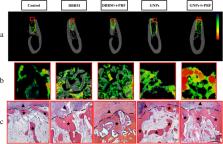
          Ridge preservation is universally acknowledged as the conventional method for the post-extraction healing yet there are no standard materials for the ideal healing outcome. Herein, a composite gel comprising gelatin nanoparticles (GNPs) and injectable platelet-rich-fibrin (i-PRF) as the potential candidate for extracted socket healing is introduced. The combination of GNPs and i-PRF not only possesses favorable mechanical properties to withstand external force but also accelerate the blood clotting time significantly. In addition, six beagle dogs were adopted to assess the angiogenic and osteogenic capacity of GNPs+i-PRF gel in vivo. The GNPs+i-PRF gel significantly produced the most blood vessels area, woven bone and low osteoclast activity in extracted sockets at 2 weeks postoperation and remarkably generated corticalization on the alveolar ridge crest at 8 weeks postoperation according to histological results. Therefore, GNPs+i-PRF gel can be recommended as the candidate grafting material regarding ridge preservation for its cost effectiveness, excellent biocompatibility, facilitation of blood clotting and favorable capacity of promoting angiogenesis and osteogenesis.

          Supplementary Information

          The online version contains supplementary material available at 10.1186/s13036-021-00271-8.

          Related collections

          Most cited references57

          • Record: found
          • Abstract: not found
          • Article: not found

          Animal research: reporting in vivo experiments: the ARRIVE guidelines.

            Bookmark
            • Record: found
            • Abstract: found
            • Article: not found

            Osteoinduction, osteoconduction and osseointegration.

            Osteoinduction is the process by which osteogenesis is induced. It is a phenomenon regularly seen in any type of bone healing process. Osteoinduction implies the recruitment of immature cells and the stimulation of these cells to develop into preosteoblasts. In a bone healing situation such as a fracture, the majority of bone healing is dependent on osteoinduction. Osteoconduction means that bone grows on a surface. This phenomenon is regularly seen in the case of bone implants. Implant materials of low biocompatibility such as copper, silver and bone cement shows little or no osteoconduction. Osseointegration is the stable anchorage of an implant achieved by direct bone-to-implant contact. In craniofacial implantology, this mode of anchorage is the only one for which high success rates have been reported. Osseointegration is possible in other parts of the body, but its importance for the anchorage of major arthroplasties is under debate. Ingrowth of bone in a porous-coated prosthesis may or may not represent osseointegration.
              Bookmark
              • Record: found
              • Abstract: found
              • Article: not found

              Dimensional ridge alterations following tooth extraction. An experimental study in the dog.

              To study dimensional alterations of the alveolar ridge that occurred following tooth extraction as well as processes of bone modelling and remodelling associated with such change. Twelve mongrel dogs were included in the study. In both quadrants of the mandible incisions were made in the crevice region of the 3rd and 4th premolars. Minute buccal and lingual full thickness flaps were elevated. The four premolars were hemi-sected. The distal roots were removed. The extraction sites were covered with the mobilized gingival tissue. The extractions of the roots and the sacrifice of the dogs were staggered in such a manner that all dogs contributed with sockets representing 1, 2, 4 and 8 weeks of healing. The animals were sacrificed and tissue blocks containing the extraction socket were dissected, decalcified in EDTA, embedded in paraffin and cut in the buccal-lingual plane. The sections were stained in haematoxyline-eosine and examined in the microscope. It was demonstrated that marked dimensional alterations occurred during the first 8 weeks following the extraction of mandibular premolars. Thus, in this interval there was a marked osteoclastic activity resulting in resorption of the crestal region of both the buccal and the lingual bone wall. The reduction of the height of the walls was more pronounced at the buccal than at the lingual aspect of the extraction socket. The height reduction was accompanied by a "horizontal" bone loss that was caused by osteoclasts present in lacunae on the surface of both the buccal and the lingual bone wall. The resorption of the buccal/lingual walls of the extraction site occurred in two overlapping phases. During phase 1, the bundle bone was resorbed and replaced with woven bone. Since the crest of the buccal bone wall was comprised solely of bundle this modelling resulted in substantial vertical reduction of the buccal crest. Phase 2 included resorption that occurred from the outer surfaces of both bone walls. The reason for this additional bone loss is presently not understood. (c) Blackwell Munksgaard, 2005.
                Bookmark

                Author and article information

                Contributors
                huananwang@dlut.edu.cn
                jiping@hospital.cqmu.edu.cn
                Journal
                J Biol Eng
                J Biol Eng
                Journal of Biological Engineering
                BioMed Central (London )
                1754-1611
                21 July 2021
                21 July 2021
                2021
                : 15
                : 19
                Affiliations
                [1 ]GRID grid.203458.8, ISNI 0000 0000 8653 0555, Stomatological Hospital of Chongqing Medical University, Chongqing Key Laboratory of Oral Diseases and Biomedical Sciences, Chongqing Municipal Key Laboratory of Oral Biomedical Engineering of Higher Education, , Chongqing Medical University, ; Chongqing, 401147 P. R. China
                [2 ]GRID grid.203458.8, ISNI 0000 0000 8653 0555, Department of Pathology, , Chongqing Medical University, ; Chongqing, 401147 P. R. China
                [3 ]GRID grid.30055.33, ISNI 0000 0000 9247 7930, Key State Laboratory of Fine Chemicals, School of Bioengineering, , Dalian University of Technology, ; No.2 Linggong Road, High-tech District, Dalian, 116024 P. R. China
                [4 ]GRID grid.268099.c, ISNI 0000 0001 0348 3990, Department of Periodontics, School and Hospital of Stomatology, , Wenzhou Medical University, ; Wenzhou, 325027 Zhejiang China
                Article
                271
                10.1186/s13036-021-00271-8
                8293569
                34289877
                26e4e216-d056-412c-88dc-3bff09f63d7b
                © The Author(s) 2021

                Open AccessThis article is licensed under a Creative Commons Attribution 4.0 International License, which permits use, sharing, adaptation, distribution and reproduction in any medium or format, as long as you give appropriate credit to the original author(s) and the source, provide a link to the Creative Commons licence, and indicate if changes were made. The images or other third party material in this article are included in the article's Creative Commons licence, unless indicated otherwise in a credit line to the material. If material is not included in the article's Creative Commons licence and your intended use is not permitted by statutory regulation or exceeds the permitted use, you will need to obtain permission directly from the copyright holder. To view a copy of this licence, visit http://creativecommons.org/licenses/by/4.0/. The Creative Commons Public Domain Dedication waiver ( http://creativecommons.org/publicdomain/zero/1.0/) applies to the data made available in this article, unless otherwise stated in a credit line to the data.

                History
                : 19 April 2021
                : 16 June 2021
                Funding
                Funded by: FundRef http://dx.doi.org/10.13039/501100001809, National Natural Science Foundation of China;
                Award ID: 31871464
                Award ID: 31870957
                Award Recipient :
                Funded by: FundRef http://dx.doi.org/10.13039/501100010031, Postdoctoral Research Foundation of China;
                Award ID: 2017M622981
                Award Recipient :
                Funded by: Chongqing Postdoctoral Science Special Foundation (CN)
                Award ID: XmT2018009
                Award Recipient :
                Funded by: FundRef http://dx.doi.org/10.13039/501100012226, Fundamental Research Funds for the Central Universities;
                Award ID: DUT15RC (3)113
                Award Recipient :
                Funded by: National Natural Science Foundation of China (CN)
                Award ID: 81701031
                Award Recipient :
                Categories
                Research
                Custom metadata
                © The Author(s) 2021

                Biotechnology
                i-prf,ridge preservation,biomaterials,gelatin,dental implant
                Biotechnology
                i-prf, ridge preservation, biomaterials, gelatin, dental implant

                Comments

                Comment on this article