0
views
0
recommends
+1 Recommend
0 collections
    0
    shares
      • Record: found
      • Abstract: found
      • Article: found
      Is Open Access

      In Vivo Injection of Anti-LGI1 Antibodies into the Rodent M1 Cortex and Hippocampus Is Ineffective in Inducing Seizures

      research-article

      Read this article at

      Bookmark
          There is no author summary for this article yet. Authors can add summaries to their articles on ScienceOpen to make them more accessible to a non-specialist audience.

          Abstract

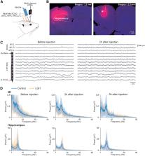
          Autoimmune encephalitis (AIE) associated with antibodies directed against the leucine-rich glioma inactivated 1 (LGI1) protein is the second most common AIE and is responsible for deleterious neocortical and limbic epileptic seizures. Previous studies demonstrated a pathogenic role of anti-LGI1 antibodies via alterations in the expression and function of Kv1 channels and AMPA receptors. However, the causal link between antibodies and epileptic seizures has never been demonstrated. Here, we attempted to determine the role of human anti-LGI1 autoantibodies in the genesis of seizures by analyzing the impact of their intracerebral injection in rodents. Acute and chronic injections were performed in rats and mice in the hippocampus and primary motor cortex, the two main brain regions affected by the disease. Acute infusion of CSF or serum IgG of anti-LGI1 AIE patients did not lead to the emergence of epileptic activities, as assessed by multisite electrophysiological recordings over a 10 h period after injection. A chronic 14 d injection, coupled with continuous video-EEG monitoring, was not more effective. Overall, these results demonstrate that acute and chronic injections of CSF or purified IgG from LGI1 patients are not able to generate epileptic activity by themselves in the different animal models tested.

          Related collections

          Most cited references39

          • Record: found
          • Abstract: found
          • Article: found
          Is Open Access

          FieldTrip: Open Source Software for Advanced Analysis of MEG, EEG, and Invasive Electrophysiological Data

          This paper describes FieldTrip, an open source software package that we developed for the analysis of MEG, EEG, and other electrophysiological data. The software is implemented as a MATLAB toolbox and includes a complete set of consistent and user-friendly high-level functions that allow experimental neuroscientists to analyze experimental data. It includes algorithms for simple and advanced analysis, such as time-frequency analysis using multitapers, source reconstruction using dipoles, distributed sources and beamformers, connectivity analysis, and nonparametric statistical permutation tests at the channel and source level. The implementation as toolbox allows the user to perform elaborate and structured analyses of large data sets using the MATLAB command line and batch scripting. Furthermore, users and developers can easily extend the functionality and implement new algorithms. The modular design facilitates the reuse in other software packages.
            Bookmark
            • Record: found
            • Abstract: found
            • Article: not found

            Human N-methyl D-aspartate receptor antibodies alter memory and behaviour in mice.

            Anti-N-methyl D-aspartate receptor (NMDAR) encephalitis is a severe neuropsychiatric disorder that associates with prominent memory and behavioural deficits. Patients' antibodies react with the N-terminal domain of the GluN1 (previously known as NR1) subunit of NMDAR causing in cultured neurons a selective and reversible internalization of cell-surface receptors. These effects and the frequent response to immunotherapy have suggested an antibody-mediated pathogenesis, but to date there is no animal model showing that patients' antibodies cause memory and behavioural deficits. To develop such a model, C57BL6/J mice underwent placement of ventricular catheters connected to osmotic pumps that delivered a continuous infusion of patients' or control cerebrospinal fluid (flow rate 0.25 µl/h, 14 days). During and after the infusion period standardized tests were applied, including tasks to assess memory (novel object recognition in open field and V-maze paradigms), anhedonic behaviours (sucrose preference test), depressive-like behaviours (tail suspension, forced swimming tests), anxiety (black and white, elevated plus maze tests), aggressiveness (resident-intruder test), and locomotor activity (horizontal and vertical). Animals sacrificed at Days 5, 13, 18, 26 and 46 were examined for brain-bound antibodies and the antibody effects on total and synaptic NMDAR clusters and protein concentration using confocal microscopy and immunoblot analysis. These experiments showed that animals infused with patients' cerebrospinal fluid, but not control cerebrospinal fluid, developed progressive memory deficits, and anhedonic and depressive-like behaviours, without affecting other behavioural or locomotor tasks. Memory deficits gradually worsened until Day 18 (4 days after the infusion stopped) and all symptoms resolved over the next week. Accompanying brain tissue studies showed progressive increase of brain-bound human antibodies, predominantly in the hippocampus (maximal on Days 13-18), that after acid extraction and characterization with GluN1-expressing human embryonic kidney cells were confirmed to be against the NMDAR. Confocal microscopy and immunoblot analysis of the hippocampus showed progressive decrease of the density of total and synaptic NMDAR clusters and total NMDAR protein concentration (maximal on Day 18), without affecting the post-synaptic density protein 95 (PSD95) and α-amino-3-hydroxy-5-methyl-4-isoxazolepropionic acid (AMPA) receptors. These effects occurred in parallel with memory and other behavioural deficits and gradually improved after Day 18, with reversibility of symptoms accompanied by a decrease of brain-bound antibodies and restoration of NMDAR levels. Overall, these findings establish a link between memory and behavioural deficits and antibody-mediated reduction of NMDAR, provide the biological basis by which removal of antibodies and antibody-producing cells improve neurological function, and offer a model for testing experimental therapies in this and similar disorders. © The Author (2014). Published by Oxford University Press on behalf of the Guarantors of Brain. All rights reserved. For Permissions, please email: journals.permissions@oup.com.
              Bookmark
              • Record: found
              • Abstract: found
              • Article: not found

              Autoantibodies to Synaptic Receptors and Neuronal Cell Surface Proteins in Autoimmune Diseases of the Central Nervous System.

              Investigations in the last 10 years have revealed a new category of neurological diseases mediated by antibodies against cell surface and synaptic proteins. There are currently 16 such diseases all characterized by autoantibodies against neuronal proteins involved in synaptic signaling and plasticity. In clinical practice these findings have changed the diagnostic and treatment approach to potentially lethal, but now treatable, neurological and psychiatric syndromes previously considered idiopathic or not even suspected to be immune-mediated. Studies show that patients' antibodies can impair the surface dynamics of the target receptors eliminating them from synapses (e.g., NMDA receptor), block the function of the antigens without changing their synaptic density (e.g., GABAb receptor), interfere with synaptic protein-protein interactions (LGI1, Caspr2), alter synapse formation (e.g., neurexin-3α), or by unclear mechanisms associate to a new form of tauopathy (IgLON5). Here we first trace the process of discovery of these diseases, describing the triggers and symptoms related to each autoantigen, and then review in detail the structural and functional alterations caused by the autoantibodies with special emphasis in those (NMDA receptor, amphiphysin) that have been modeled in animals.
                Bookmark

                Author and article information

                Journal
                eNeuro
                eNeuro
                eneuro
                eNeuro
                eNeuro
                Society for Neuroscience
                2373-2822
                27 February 2023
                13 March 2023
                March 2023
                : 10
                : 3
                : ENEURO.0267-22.2023
                Affiliations
                [1 ]Sorbonne Université, Institut du Cerveau - Paris Brain Institute - ICM, Inserm, CNRS, APHP, Hôpital de la Pitié-Salpêtriére, 75013 Paris, France
                [2 ]AP-HP, Hôpital de la Pitié-Salpêtriére, DMU Neurosciences 6, Epilepsy Unit and Clinical Neurophysiology Department, 75013 Paris, France
                [3 ]Center of Reference for Rare Epilepsies, APHP, Hôpital de la Pitié-Salpêtrière, 47 Boulevard de l’Hôpital, 75013 Paris, France
                Author notes

                V.N. reports personal fees from UCB, Liva Nova, and EISAI, outside of the submitted work. The authors declare no other competing financial interests.

                Author contributions: P.B., S.M., S.C., and V.N. designed research; P.B. and D.R. performed research; P.B. contributed unpublished reagents/analytic tools; P.B. analyzed data; P.B., S.M., S.C., and V.N. wrote the paper.

                This research was supported by Agence Nationale de la Recherche, “Investissements d’avenir” Program Grants ANR-10-IAIHU-06 and ANR-11-INBS-0011-NeurATRIS; Fondation pour la Recherche Médicale, Grant FDT202012010523; and Fondation Assitance Publique-Hôpitaux de Paris (EPIRES, Marie Laure PLV Merchandising).

                Correspondence should be addressed to Vincent Navarro at vincent.navarro@ 123456aphp.fr .
                Author information
                https://orcid.org/0000-0002-8714-5784
                Article
                eN-NRS-0267-22
                10.1523/ENEURO.0267-22.2023
                10012326
                36849262
                27e14a90-3135-4f9a-be3d-a36d263667b3
                Copyright © 2023 Baudin et al.

                This is an open-access article distributed under the terms of the Creative Commons Attribution 4.0 International license, which permits unrestricted use, distribution and reproduction in any medium provided that the original work is properly attributed.

                History
                : 1 July 2022
                : 22 December 2022
                : 2 February 2023
                Page count
                Figures: 2, Tables: 3, Equations: 0, References: 39, Pages: 9, Words: 00
                Funding
                Funded by: Agence Nationale de la Recherche (ANR), doi 10.13039/501100001665;
                Award ID: ANR-10IAIHU-06
                Award ID: ANR-11-INBS-011-NeurATRIS
                Funded by: Fondation pour la Recherche Médicale (FRM), doi 10.13039/501100002915;
                Award ID: FDT202012010523
                Funded by: Fondation Assistance Publique-Hôpitaux de Paris
                Award ID: EPIRES
                Award ID: Marie Laure PLV Merchandising
                Categories
                3
                Research Article: Negative Results
                Disorders of the Nervous System
                Custom metadata
                March 2023

                autoimmune encephalitis,electrophysiology,epilepsy,lgi1,video-eeg

                Comments

                Comment on this article