151
views
0
recommends
+1 Recommend
0 collections
    0
    shares
      • Record: found
      • Abstract: found
      • Article: not found

      Multi-task connectivity reveals flexible hubs for adaptive task control

      research-article

      Read this article at

      ScienceOpenPublisherPMC
      Bookmark
          There is no author summary for this article yet. Authors can add summaries to their articles on ScienceOpen to make them more accessible to a non-specialist audience.

          Abstract

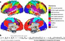
          Extensive evidence suggests the human ability to adaptively implement a wide variety of tasks is preferentially due to the operation of a fronto-parietal brain network. We hypothesized that this network’s adaptability is made possible by ‘flexible hubs’ – brain regions that rapidly update their pattern of global functional connectivity according to task demands. We utilized recent advances in characterizing brain network organization and dynamics to identify mechanisms consistent with the flexible hub theory. We found that the fronto-parietal network’s brain-wide functional connectivity pattern shifted more than other networks’ across a variety of task states, and that these connectivity patterns could be used to identify the current task. Further, these patterns were consistent across practiced and novel tasks, suggesting reuse of flexible hub connectivity patterns facilitates adaptive (novel) task performance. Together, these findings support a central role for fronto-parietal flexible hubs in cognitive control and adaptive implementation of task demands generally.

          Related collections

          Most cited references61

          • Record: found
          • Abstract: found
          • Article: not found

          An automated labeling system for subdividing the human cerebral cortex on MRI scans into gyral based regions of interest.

          In this study, we have assessed the validity and reliability of an automated labeling system that we have developed for subdividing the human cerebral cortex on magnetic resonance images into gyral based regions of interest (ROIs). Using a dataset of 40 MRI scans we manually identified 34 cortical ROIs in each of the individual hemispheres. This information was then encoded in the form of an atlas that was utilized to automatically label ROIs. To examine the validity, as well as the intra- and inter-rater reliability of the automated system, we used both intraclass correlation coefficients (ICC), and a new method known as mean distance maps, to assess the degree of mismatch between the manual and the automated sets of ROIs. When compared with the manual ROIs, the automated ROIs were highly accurate, with an average ICC of 0.835 across all of the ROIs, and a mean distance error of less than 1 mm. Intra- and inter-rater comparisons yielded little to no difference between the sets of ROIs. These findings suggest that the automated method we have developed for subdividing the human cerebral cortex into standard gyral-based neuroanatomical regions is both anatomically valid and reliable. This method may be useful for both morphometric and functional studies of the cerebral cortex as well as for clinical investigations aimed at tracking the evolution of disease-induced changes over time, including clinical trials in which MRI-based measures are used to examine response to treatment.
            Bookmark
            • Record: found
            • Abstract: found
            • Article: not found

            LIBSVM: A library for support vector machines

            LIBSVM is a library for Support Vector Machines (SVMs). We have been actively developing this package since the year 2000. The goal is to help users to easily apply SVM to their applications. LIBSVM has gained wide popularity in machine learning and many other areas. In this article, we present all implementation details of LIBSVM. Issues such as solving SVM optimization problems theoretical convergence multiclass classification probability estimates and parameter selection are discussed in detail.
              Bookmark
              • Record: found
              • Abstract: found
              • Article: not found

              Complex network measures of brain connectivity: uses and interpretations.

              Brain connectivity datasets comprise networks of brain regions connected by anatomical tracts or by functional associations. Complex network analysis-a new multidisciplinary approach to the study of complex systems-aims to characterize these brain networks with a small number of neurobiologically meaningful and easily computable measures. In this article, we discuss construction of brain networks from connectivity data and describe the most commonly used network measures of structural and functional connectivity. We describe measures that variously detect functional integration and segregation, quantify centrality of individual brain regions or pathways, characterize patterns of local anatomical circuitry, and test resilience of networks to insult. We discuss the issues surrounding comparison of structural and functional network connectivity, as well as comparison of networks across subjects. Finally, we describe a Matlab toolbox (http://www.brain-connectivity-toolbox.net) accompanying this article and containing a collection of complex network measures and large-scale neuroanatomical connectivity datasets. Copyright (c) 2009 Elsevier Inc. All rights reserved.
                Bookmark

                Author and article information

                Journal
                9809671
                21092
                Nat Neurosci
                Nat. Neurosci.
                Nature neuroscience
                1097-6256
                1546-1726
                22 July 2013
                28 July 2013
                September 2013
                01 March 2014
                : 16
                : 9
                : 1348-1355
                Affiliations
                [1 ]Department of Psychology, Washington University in St. Louis, MO, USA 63130
                [2 ]Department of Psychology, University of Denver, Denver, CO, USA 80208
                [3 ]Department of Neurology, Washington University in St. Louis, MO, USA 63130
                [4 ]Department of Psychology, University of Ljubljana, Slovenia
                [5 ]Department of Psychiatry, Yale University, New Haven, CT 06511
                Author notes
                Author contact info: Michael W. Cole, mwcole@ 123456mwcole.net , One Brookings Dr., Campus Box 1125, St. Louis, MO 63130
                Article
                NIHMS497578
                10.1038/nn.3470
                3758404
                23892552
                27f28511-ad14-4346-997a-b85da12c105b

                Users may view, print, copy, download and text and data- mine the content in such documents, for the purposes of academic research, subject always to the full Conditions of use: http://www.nature.com/authors/editorial_policies/license.html#terms

                History
                Funding
                Funded by: National Institute of Mental Health : NIMH
                Award ID: K99 MH096801 || MH
                Categories
                Article

                Neurosciences
                Neurosciences

                Comments

                Comment on this article