37
views
0
recommends
+1 Recommend
0 collections
    0
    shares
      • Record: found
      • Abstract: found
      • Article: not found

      Depolarization-induced Calcium Responses in Sympathetic Neurons: Relative Contributions from Ca 2+ Entry, Extrusion, ER/Mitochondrial Ca 2+ Uptake and Release, and Ca 2+ Buffering

      research-article

      Read this article at

      ScienceOpenPublisherPMC
      Bookmark
          There is no author summary for this article yet. Authors can add summaries to their articles on ScienceOpen to make them more accessible to a non-specialist audience.

          Abstract

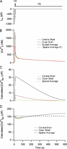
          Many models have been developed to account for stimulus-evoked [Ca 2+] responses, but few address how responses elicited in specific cell types are defined by the Ca 2+ transport and buffering systems that operate in the same cells. In this study, we extend previous modeling studies by linking the time course of stimulus-evoked [Ca 2+] responses to the underlying Ca 2+ transport and buffering systems. Depolarization-evoked [Ca 2+] i responses were studied in sympathetic neurons under voltage clamp, asking how response kinetics are defined by the Ca 2+ handling systems expressed in these cells. We investigated five cases of increasing complexity, comparing observed and calculated responses deduced from measured Ca 2+ handling properties. In Case 1, [Ca 2+] i responses were elicited by small Ca 2+ currents while Ca 2+ transport by internal stores was inhibited, leaving plasma membrane Ca 2+ extrusion intact. In Case 2, responses to the same stimuli were measured while mitochondrial Ca 2+ uptake was active. In Case 3, responses were elicited as in Case 2 but with larger Ca 2+ currents that produce larger and faster [Ca 2+] i elevations. Case 4 included the mitochondrial Na/Ca exchanger. Finally, Case 5 included ER Ca 2+ uptake and release pathways. We found that [Ca 2+] i responses elicited by weak stimuli (Cases 1 and 2) could be quantitatively reconstructed using a spatially uniform model incorporating the measured properties of Ca 2+ entry, removal, and buffering. Responses to strong depolarization (Case 3) could not be described by this model, but were consistent with a diffusion model incorporating the same Ca 2+ transport and buffering descriptions, as long as endogenous buffers have low mobility, leading to steep radial [Ca 2+] i gradients and spatially nonuniform Ca 2+ loading by mitochondria. When extended to include mitochondrial Ca 2+ release (Case 4) and ER Ca 2+ transport (Case 5), the diffusion model could also account for previous measurements of stimulus-evoked changes in total mitochondrial and ER Ca concentration.

          Related collections

          Most cited references51

          • Record: found
          • Abstract: found
          • Article: not found

          Generation, control, and processing of cellular calcium signals.

          In the course of evolution, Ca2+ has emerged as the most versatile intracellular messenger. Its concentration within cells is controlled by reversible binding to specific classes of proteins that act as Ca2+ sensors to decode its information before passing it on to targets. The decoding operation is based on specific conformational changes in the sensor proteins. Other proteins intrinsic to membranes simply control Ca2+ concentration without processing its message, by transporting it across membrane boundaries. They are located in the plasma membrane and in the membranes of the organelles (the endo(sarco)plasmic reticulum, the mitochondria, the nuclear envelope), which play distinctive roles in the cellular homeostasis of Ca2+. Ca2+ is an ambivalent signaling agent. It carries information to virtually all processes important to cell life (e.g., it couples excitation to contraction, secretion, gene transcription, and controls enzyme activity through protein phosphorylation-dephosphorylation), but also transmits signals that promote the programmed demise of cells. When escaping control, Ca2+ also precipitates toxic cell death.
            Bookmark
            • Record: found
            • Abstract: found
            • Article: not found

            Calcium gradients and buffers in bovine chromaffin cells.

            1. Digital imaging and photometry were used in conjunction with the fluorescent Ca2+ indicator, Fura-2, to examine intracellular Ca2+ signals produced by depolarization of single adrenal chromaffin cells. 2. Depolarization with a patch pipette produced radial gradients of Ca2+ within the cell, with Ca2+ concentration highest in the vicinity of the plasma membrane. These gradients dissipated within a few hundred milliseconds when the voltage-gated Ca2+ channels were closed. 3. Dialysis of Fura-2 into the chromaffin cell caused concentration-dependent changes in the depolarization-induced Ca2+ signal, decreasing its magnitude and slowing its recovery time course. These changes were used to estimate the properties of the endogenous cytoplasmic Ca2+ buffer with which Fura-2 competes for Ca2+. 4. The spatially averaged Fura-2 signal was well described by a model assuming fast competition between Fura-2 and an endogenous buffer on a millisecond time scale. Retrieval of calcium by pumps and slow buffers occurs on a seconds-long time scale. No temporal changes indicative of buffers with intermediate kinetics could be detected. 5. Two independent estimates of the capacity of the fast endogenous Ca2+ buffer suggest that 98-99% of the Ca2+ entering the cell normally is taken up by this buffer. This buffer appears to be immobile, because it does not wash out of the cell during dialysis. It has a low affinity for Ca2+ ions, because it does not saturate with 1 microM-Ca2+ inside the cell. 6. The low capacity, affinity and mobility of the endogenous Ca2+ buffer makes it possible for relatively small amounts of exogenous Ca2+ buffers, such as Fura-2, to exert a significant influence on the characteristics of the Ca2+ concentration signal as measured by fluorescence ratios. On the other hand, even at moderate Fura-2 concentrations (0.4 mM) Fura-2 will dominate over the endogenous buffers. Under these conditions radiometric Ca2+ concentration signals are largely attenuated, but absolute fluorescence changes (at 390 nm) accurately reflect calcium fluxes.
              Bookmark
              • Record: found
              • Abstract: found
              • Article: not found

              Minimal model for signal-induced Ca2+ oscillations and for their frequency encoding through protein phosphorylation.

              In a variety of cells, hormonal or neurotransmitter signals elicit a train of intracellular Ca2+ spikes. The analysis of a minimal model based on Ca2(+)-induced Ca2+ release from intracellular stores shows how sustained oscillations of cytosolic Ca2+ may develop as a result of a rise in inositol 1,4,5-trisphosphate (InsP3) triggered by external stimulation. This rise elicits the release of a certain amount of Ca2+ from an InsP3-sensitive intracellular store. The subsequent rise in cytosolic Ca2+ in turn triggers the release of Ca2+ from a second store insensitive to InsP3. In contrast to the model proposed by Meyer and Stryer [Meyer, T. & Stryer, L. (1988) Proc. Natl. Acad. Sci. USA 85, 5051-5055], the present model, which contains only two variables, predicts the occurrence of periodic Ca2+ spikes in the absence of InsP3 oscillations. Such results indicate that repetitive Ca2+ spikes evoked by external stimuli do not necessarily require the concomitant, periodic variation of InsP3. The model is closely related to that proposed by Kuba and Takeshita [Kuba, K. & Takeshita, S. (1981) J. Theor. Biol. 93, 1009-1031] for Ca2+ oscillations in sympathetic neurones, based on Ca2(+)-induced Ca2+ release. We extend their results by showing the minimal conditions in which the latter process gives rise to periodic behavior and take into account the role of the rise in InsP3 caused by external stimulation. The analysis further shows how signal-induced Ca2+ oscillations might be effectively encoded in terms of their frequency through the phosphorylation of a cellular substrate by a protein kinase activated by cytosolic Ca2+.
                Bookmark

                Author and article information

                Journal
                J Gen Physiol
                The Journal of General Physiology
                The Rockefeller University Press
                0022-1295
                1540-7748
                January 2007
                : 129
                : 1
                : 29-56
                Affiliations
                [1 ]Department of Neurosciences, Case Western Reserve University, Cleveland, OH 44106
                [2 ]Department of Mathematics, University of Auckland, Auckland, New Zealand
                Author notes

                Correspondence to David Friel: ddf2@ 123456case.edu

                Article
                200609660
                10.1085/jgp.200609660
                2151609
                17190902
                27fe3654-043b-464d-a947-b66128dd80d1
                Copyright © 2007, The Rockefeller University Press
                History
                : 7 September 2006
                : 8 December 2006
                Categories
                Articles
                Article

                Anatomy & Physiology
                Anatomy & Physiology

                Comments

                Comment on this article