17
views
0
recommends
+1 Recommend
0 collections
    0
    shares
      • Record: found
      • Abstract: found
      • Article: found
      Is Open Access

      Comparison of immunohistochemical expression of CD10 in keratocystic odontogenic tumor and ameloblastoma

      research-article

      Read this article at

      Bookmark
          There is no author summary for this article yet. Authors can add summaries to their articles on ScienceOpen to make them more accessible to a non-specialist audience.

          Abstract

          Background:

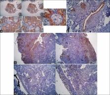
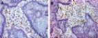
          Odontogenic keratocyst (OKC), also called keratocystic odontogenic tumor (KCOT), is a developmental lesion which should be carefully monitored and it exhibits development mechanisms and biologic behaviors different from those of other more common lesions such as dentigerous and radicular cysts. CD10 antigen is a cell surface metalloendopeptidase, which inactivates various peptides that are physiologically active. Studies have shown that increase in the expression of CD10 in the stromal cells helps the progression of the tumor. Ameloblastoma (AB) is a local invasive tumor and given the role of supporting connective tissue stroma in the aggression and progression. The aim of the present study was to comparatively evaluate the expression of CD10 in the connective tissue stroma of AB and OKC as a KCOT.

          Materials and Methods:

          In this retrospective, cross-sectional study, 14 paraffin blocks of KCOT and 9 of AB (7 multicystic and 2 unicystic) were evaluated with CD10 immunohistochemical expression in the connective tissue stroma of AB and the connective tissue wall of KCOT. The data were analyzed with Fisher's exact test ( P < 0.05).

          Results:

          In 8 samples of 9 AB and in 13 samples of 14 KOT lesions, expression of CD10 was shown. Fisher's exact test did not reveal any significant differences between these two lesions in the expression of CD10 ( P = 0.64).

          Conclusion:

          The results of this study propose that high expression rate of CD10 might be one of the reasons for the aggressive behavior of AB and high recurrence rate of OKC and reinforce the classification of OKC as an odontogenic tumor.

          Related collections

          Most cited references25

          • Record: found
          • Abstract: found
          • Article: not found

          The CD10 enzyme is a key player to identify and regulate human mammary stem cells.

          The major components of the mammary ductal tree are an inner layer of luminal cells, an outer layer of myoepithelial cells, and a basement membrane that separates the ducts from the underlying stroma. Cells in the outer layer express CD10, a zinc-dependent metalloprotease that regulates the growth of the ductal tree during mammary gland development. To define the steps in the human mammary lineage at which CD10 acts, we have developed an in vitro assay for human mammary lineage progression. We show that sorting for CD10 and EpCAM cleanly separates progenitors from differentiated luminal cells and that the CD10-high EpCAM-low population is enriched for early common progenitor and mammosphere-forming cells. We also show that sorting for CD10 enriches sphere-forming cells from other tissue types, suggesting that it may provide a simple tool to identify stem or progenitor populations in tissues for which lineage studies are not currently possible. We demonstrate that the protease activity of CD10 and the adhesion function of beta1-integrin are required to prevent differentiation of mammary progenitors. Taken together, our data suggest that integrin-mediated contact with the basement membrane and cleavage of signaling factors by CD10 are key elements in the niche that maintains the progenitor and stem cell pools in the mammary lineage.
            Bookmark
            • Record: found
            • Abstract: found
            • Article: not found

            Expression of CD10 by stromal cells during colorectal tumor development.

            CD10 is a cell surface metalloprotease expressed by a variety of normal cell types, including lymphoid precursor cells, germinal center B lymphocytes, and some epithelial cells. We noticed that stromal cells of some cancers are positive for CD10. In this study, we investigated the role of CD10 produced by the stromal cells of colorectal neoplasms in the progression of colorectal neoplasms. Immunohistochemical examination of CD10 and p53 was performed in 169 colorectal epithelial neoplasms representing various stages of carcinogenesis. The results were correlated with the morphologic characteristics of the neoplasms. There was no expression of CD10 in the stromal cells of normal colorectal tissue. CD10-positive stromal cells were present adjacent to the tumor cells in 16 of 73 adenomas with mild or moderate dysplasia. More frequent expression of CD10 by the stromal cells was detected in adenomas with severe dysplasia (12 of 17), intramucosal carcinomas (10 of 16), and invasive carcinomas (50 of 63) than in adenomas with mild or moderate dysplasia (P 10% of the stromal cells was detected only within the area of the invasive growth front of invasive carcinomas, not in adenomas and in only 1 of the intramucosal carcinomas. The difference between invasive and non invasive tumors was significant (P < 0.0001). The stromal expression of CD10 was significantly associated with the accumulation of p53 and a larger tumor size. These results indicate that CD10 expression is an integral part of colorectal carcinogenesis. CD10 expression seems to contribute to the invasion and thus probably facilitates metastasis. Copyright 2002, Elsevier Science (USA). All rights reserved.
              Bookmark
              • Record: found
              • Abstract: found
              • Article: found
              Is Open Access

              EGFR, CD10 and proliferation marker Ki67 expression in ameloblastoma: possible role in local recurrence

              Background Ameloblastoma is an odontogenic neoplasm characterized by local invasiveness and tendency towards recurrence. Aims Studying the role played by EGFR, CD10 and Ki67 in the recurrence of ameloblastoma. Methods This study was carried out on 22 retrospective cases of mandibular ameloblastoma from the period from Jan 2002 to Jan 2008 with follow up period until Jan 2011 (3 to 8 years follow up peroid). Archival materials were obtained from pathology department, Mansoura university. Paraffin sections of tumor tissue from all cases were submitted for routine H&E stains and immunohistochemistry using EGFR, CD10 and Ki67 monoclonal antibodies. Statistical analysis using of clinical data for all patients, tumor type, EGFR, CD10 and Ki67 expression in relation to recurrence were evaluated. Results Among the 22 cases, 10 cases were males and 12 were females with sex ratio 1:1.2. Age ranged from 34 to 59 years old with a mean age 44.18 year. Five cases showed local recurrence within studied period and proved by biopsy. No statistically significant relation was found between local recurrence and patient age, tumor size, tumor type, EGFR expression. There was a significant relation between CD10 expression as well as Ki67 labelling index and recurrence (P value = 0.003, 0.000 respectively). Conclusion Evaluation of CD10 and Ki67 status together with conventional histological evaluation can help in providing more information about the biologic behavior of the tumor, while EGFR could be a target of an expanding class of anticancer therapies. Since ameloblastomas are EGFR-positive tumors, anti-EGFR agents could be considered to reduce the size of large tumors and to treat unresectable tumors that are in close proximity to vital structures. Virtual Slides The virtual slide(s) for this article can be found here: http://www.diagnosticpathology.diagnomx.eu/vs/1902106905645651
                Bookmark

                Author and article information

                Journal
                Dent Res J (Isfahan)
                Dent Res J (Isfahan)
                DRJ
                Dental Research Journal
                Medknow Publications & Media Pvt Ltd (India )
                1735-3327
                2008-0255
                Mar-Apr 2016
                : 13
                : 2
                : 110-116
                Affiliations
                [1 ]Department of Orthodontics, Faculty of Dentistry, Ahvaz Jundishapur University of Medical Sciences, Ahvaz, Iran
                [2 ]Torabinejad Dental Research Center and Department of Oral and Maxillofacial Pathology, School of Dentistry, Isfahan University of Medical Sciences, Isfahan, Iran
                Author notes
                Address for correspondence: Dr. Forooz Keshani, Department of Oral and Maxillofacial Pathology, Torabinejad Dental Research Center, School of Dentistry, Isfahan University of Medical Sciences, Isfahan, Iran. E-mail: kforuz@ 123456yahoo.com
                Article
                DRJ-13-110
                4810907
                27076824
                2858e120-abf9-49f6-a4ab-76462055e5c0
                Copyright: © Dental Research Journal

                This is an open access article distributed under the terms of the Creative Commons Attribution NonCommercial ShareAlike 3.0 License, which allows others to remix, tweak, and build upon the work non commercially, as long as the author is credited and the new creations are licensed under the identical terms.

                History
                : April 2015
                : November 2015
                Categories
                Original Article

                Dentistry
                ameloblastoma,cd10,keratocystic,odontogenic tumor,odontogenic keratocyst
                Dentistry
                ameloblastoma, cd10, keratocystic, odontogenic tumor, odontogenic keratocyst

                Comments

                Comment on this article