16
views
0
recommends
+1 Recommend
0 collections
    0
    shares
      • Record: found
      • Abstract: found
      • Article: found
      Is Open Access

      DHPR activation underlies SR Ca 2+ release induced by osmotic stress in isolated rat skeletal muscle fibers

      research-article

      Read this article at

      Bookmark
          There is no author summary for this article yet. Authors can add summaries to their articles on ScienceOpen to make them more accessible to a non-specialist audience.

          Abstract

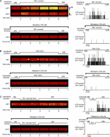
          Changes in skeletal muscle volume induce localized sarcoplasmic reticulum (SR) Ca 2+ release (LCR) events, which are sustained for many minutes, suggesting a possible signaling role in plasticity or pathology. However, the mechanism by which cell volume influences SR Ca 2+ release is uncertain. In the present study, rat flexor digitorum brevis fibers were superfused with isoosmotic Tyrode's solution before exposure to either hyperosmotic (404 mOsm) or hypoosmotic (254 mOsm) solutions, and the effects on cell volume, membrane potential (E m), and intracellular Ca 2+ ([Ca 2+] i) were determined. To allow comparison with previous studies, solutions were made hyperosmotic by the addition of sugars or divalent cations, or they were made hypoosmotic by reducing [NaCl] o. All hyperosmotic solutions induced a sustained decrease in cell volume, which was accompanied by membrane depolarization (by 14–18 mV; n = 40) and SR Ca 2+ release. However, sugar solutions caused a global increase in [Ca 2+] i, whereas solutions made hyperosmotic by the addition of divalent cations only induced LCR. Decreasing osmolarity induced an increase in cell volume and a negative shift in E m (by 15.04 ± 1.85 mV; n = 8), whereas [Ca 2+] i was unaffected. However, on return to the isoosmotic solution, restoration of cell volume and E m was associated with LCR. Both global and localized SR Ca 2+ release were abolished by the dihydropyridine receptor inhibitor nifedipine by sustained depolarization of the sarcolemmal or by the addition of the ryanodine receptor 1 inhibitor tetracaine. Inhibitors of the Na-K-2Cl (NKCC) cotransporter markedly inhibited the depolarization associated with hyperosmotic shrinkage and the associated SR Ca 2+ release. These findings suggest (1) that the depolarization that accompanies a decrease in cell volume is the primary event leading to SR Ca 2+ release, and (2) that volume-dependent regulation of the NKCC cotransporter contributes to the observed changes in E m. The differing effects of the osmotic agents can be explained by the screening of fixed charges by divalent ions.

          Related collections

          Most cited references35

          • Record: found
          • Abstract: found
          • Article: not found

          Dynamics and consequences of potassium shifts in skeletal muscle and heart during exercise.

          Since it became clear that K(+) shifts with exercise are extensive and can cause more than a doubling of the extracellular [K(+)] ([K(+)](s)) as reviewed here, it has been suggested that these shifts may cause fatigue through the effect on muscle excitability and action potentials (AP). The cause of the K(+) shifts is a transient or long-lasting mismatch between outward repolarizing K(+) currents and K(+) influx carried by the Na(+)-K(+) pump. Several factors modify the effect of raised [K(+)](s) during exercise on membrane potential (E(m)) and force production. 1) Membrane conductance to K(+) is variable and controlled by various K(+) channels. Low relative K(+) conductance will reduce the contribution of [K(+)](s) to the E(m). In addition, high Cl(-) conductance may stabilize the E(m) during brief periods of large K(+) shifts. 2) The Na(+)-K(+) pump contributes with a hyperpolarizing current. 3) Cell swelling accompanies muscle contractions especially in fast-twitch muscle, although little in the heart. This will contribute considerably to the lowering of intracellular [K(+)] ([K(+)](c)) and will attenuate the exercise-induced rise of intracellular [Na(+)] ([Na(+)](c)). 4) The rise of [Na(+)](c) is sufficient to activate the Na(+)-K(+) pump to completely compensate increased K(+) release in the heart, yet not in skeletal muscle. In skeletal muscle there is strong evidence for control of pump activity not only through hormones, but through a hitherto unidentified mechanism. 5) Ionic shifts within the skeletal muscle t tubules and in the heart in extracellular clefts may markedly affect excitation-contraction coupling. 6) Age and state of training together with nutritional state modify muscle K(+) content and the abundance of Na(+)-K(+) pumps. We conclude that despite modifying factors coming into play during muscle activity, the K(+) shifts with high-intensity exercise may contribute substantially to fatigue in skeletal muscle, whereas in the heart, except during ischemia, the K(+) balance is controlled much more effectively.
            Bookmark
            • Record: found
            • Abstract: found
            • Article: not found

            Water and ion shifts in skeletal muscle of humans with intense dynamic knee extension.

            Six subjects performed one-legged dynamic knee-extension. Blood samples were drawn from the femoral artery and vein, and muscle biopsies were obtained from the quadriceps muscle. Leg blood flow was measured by the thermodilution technique, and 3H-inulin was infused for determination of extra- and intracellular muscle water shifts. During the submaximal work load (S) muscle lactate increased, whereas muscle pH remained almost constant; after maximal exercise (M) the values markedly increased for lactate and decreased for pH. Except for a release of lactate from the exercising muscles, K was continuously released throughout S, and this release increased during M. Immediately when the muscles relaxed, the K release was converted to a K re-uptake. The calculated K loss, based on v- a and flow values, agreed with the decrease in muscle K content from 458 mmol/kg dw at rest to 414 mmol/kg dw at exhaustion (P less than 0.05), as analyzed on the muscle biopsies. Muscle water content increased during S mainly because of an increased extracellular H2O, whereas during M the largest increase occurred in intracellular H2O (H2Oi). Because of the simultaneous K loss and H2Oi increase in the exercising muscle the intracellular [K] was calculated to decrease from 165 mM at rest to 129 mM at exhaustion. This decrease and an increase in extracellular [K] from 4.5 mM at rest to greater than 6.0 mM at exhaustion affects the muscle membrane excitability. Muscle fatigue may thus not only be caused by changes within the cell, affecting energy metabolism or actin-myosin reaction, but may be located at the membrane protecting the cell against overload.
              Bookmark
              • Record: found
              • Abstract: found
              • Article: not found

              Muscle aging is associated with compromised Ca2+ spark signaling and segregated intracellular Ca2+ release

              Reduced homeostatic capacity for intracellular Ca2+ ([Ca2+]i) movement may underlie the progression of sarcopenia and contractile dysfunction during muscle aging. We report two alterations to Ca2+ homeostasis in skeletal muscle that are associated with aging. Ca2+ sparks, which are the elemental units of Ca2+ release from sarcoplasmic reticulum, are silent under resting conditions in young muscle, yet activate in a dynamic manner upon deformation of membrane structures. The dynamic nature of Ca2+ sparks appears to be lost in aged skeletal muscle. Using repetitive voltage stimulation on isolated muscle preparations, we identify a segregated [Ca2+]i reserve that uncouples from the normal excitation–contraction process in aged skeletal muscle. Similar phenotypes are observed in adolescent muscle null for a synaptophysin-family protein named mitsugumin-29 (MG29) that is involved in maintenance of muscle membrane ultrastructure and Ca2+ signaling. This finding, coupled with decreased expression of MG29 in aged skeletal muscle, suggests that MG29 expression is important in maintaining skeletal muscle Ca2+ homeostasis during aging.
                Bookmark

                Author and article information

                Journal
                J Gen Physiol
                J. Gen. Physiol
                jgp
                The Journal of General Physiology
                The Rockefeller University Press
                0022-1295
                1540-7748
                May 2009
                : 133
                : 5
                : 511-524
                Affiliations
                Institute of Membrane and Systems Biology, Faculty of Biological Sciences, University of Leeds, Leeds LS2 9JT, England, UK
                Author notes
                Correspondence to Derek S. Steele: d.steele@ 123456leeds.ac.uk
                Article
                200910191
                10.1085/jgp.200910191
                2712967
                19398777
                28bb914d-23e3-4989-8251-ff8ae24622c0
                © 2009 Pickering et al.

                This article is distributed under the terms of an Attribution–Noncommercial–Share Alike–No Mirror Sites license for the first six months after the publication date (see http://www.jgp.org/misc/terms.shtml). After six months it is available under a Creative Commons License (Attribution–Noncommercial–Share Alike 3.0 Unported license, as described at http://creativecommons.org/licenses/by-nc-sa/3.0/).

                History
                : 6 January 2009
                : 10 April 2009
                Categories
                Article

                Anatomy & Physiology
                Anatomy & Physiology

                Comments

                Comment on this article