15
views
0
recommends
+1 Recommend
0 collections
    0
    shares
      • Record: found
      • Abstract: found
      • Article: found
      Is Open Access

      Embryotoxicity assays for leached components from dental restorative materials

      research-article

      Read this article at

      Bookmark
          There is no author summary for this article yet. Authors can add summaries to their articles on ScienceOpen to make them more accessible to a non-specialist audience.

          Abstract

          Background

          Currently, there are no suitable assays available to evaluate the embryotoxicity of leached components from restorative dental materials.

          Methods

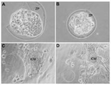
          The effect of the medium conditioned by composites and amalgam on mouse blastocysts in vitro was tested. The materials were also subcutaneously implanted, and the effect of the medium supplemented with serum from the host blood was evaluated in the embryotoxicity assay. The embryo implantation rate in the material-transplanted mothers was also evaluated.

          Results

          The results show that while the culture in media conditioned by amalgams did not affect blastocyst development, the medium conditioned by composites caused blastocyst degeneration and apoptosis. The development of blastocysts in a medium containing serum obtained from animals after transplantation was, however, without effect. Finally, inconsistent reduction in the implantation rate in transplanted mothers was observed.

          Conclusions

          In this study, we provide examples of in vitro and in vivo tests that may be used to evaluate embryotoxicity for dental materials. Our results show that leached components from our composite-material induced embryotoxicity in vitro, however, no toxicity was observed when subcutaneously implanted in vivo. This highlights the necessity of integrated in vitro and in vivo tests for valuable predictive estimation of embryotoxicity for complex materials.

          Related collections

          Most cited references17

          • Record: found
          • Abstract: found
          • Article: not found

          Identification of programmed cell death in situ via specific labeling of nuclear DNA fragmentation

          Programmed cell death (PCD) plays a key role in developmental biology and in maintenance of the steady state in continuously renewing tissues. Currently, its existence is inferred mainly from gel electrophoresis of a pooled DNA extract as PCD was shown to be associated with DNA fragmentation. Based on this observation, we describe here the development of a method for the in situ visualization of PCD at the single-cell level, while preserving tissue architecture. Conventional histological sections, pretreated with protease, were nick end labeled with biotinylated poly dU, introduced by terminal deoxy- transferase, and then stained using avidin-conjugated peroxidase. The reaction is specific, only nuclei located at positions where PCD is expected are stained. The initial screening includes: small and large intestine, epidermis, lymphoid tissues, ovary, and other organs. A detailed analysis revealed that the process is initiated at the nuclear periphery, it is relatively short (1-3 h from initiation to cell elimination) and that PCD appears in tissues in clusters. The extent of tissue-PCD revealed by this method is considerably greater than apoptosis detected by nuclear morphology, and thus opens the way for a variety of studies.
            Bookmark
            • Record: found
            • Abstract: found
            • Article: not found

            Estrogenicity of resin-based composites and sealants used in dentistry.

            We tested some resin-based composites used in dentistry for their estrogenic activity. A sealant based on bisphenol-A diglycidylether methacrylate (bis-GMA) increased cell yields, progesterone receptor expression, and pS2 secretion in human estrogen-target, serum-sensitive MCF7 breast cancer cells. Estrogenicity was due to bisphenol-A and bisphenol-A dimethacrylate, monomers found in the base paste of the dental sealant and identified by mass spectrometry. Samples of saliva from 18 subjects treated with 50 mg of a bis-GMA-based sealant applied on their molars were collected 1 hr before and after treatment. Bisphenol-A (range 90-931 micrograms) was identified only in saliva collected during a 1-hr period after treatment. The use of bis-GMA-based resins in dentistry, and particularly the use of sealants in children, appears to contribute to human exposure to xenoestrogens. Images Figure 1. A Figure 1. B Figure 2. Figure 3. A Figure 3. B Figure 4. A Figure 4. B Figure 5. A Figure 5. B Figure 6. A Figure 6. B Figure 7. A Figure 7. B Figure 8. Figure 9. Figure 10.
              Bookmark
              • Record: found
              • Abstract: not found
              • Article: not found

              Bisphenol-A: an estrogenic substance is released from polycarbonate flasks during autoclaving

                Bookmark

                Author and article information

                Journal
                Reprod Biol Endocrinol
                Reproductive Biology and Endocrinology : RB&E
                BioMed Central
                1477-7827
                2011
                6 October 2011
                : 9
                : 136
                Affiliations
                [1 ]Department of Dental Science, University of Rome "Tor Vergata", Rome, Italy
                [2 ]Department of Health Science, University of L'Aquila, L'Aquila, Italy
                [3 ]Department of Public Health and Cell Biology, Section of Histology and Embryology, University of Rome "Tor Vergata," Rome, Italy
                Article
                1477-7827-9-136
                10.1186/1477-7827-9-136
                3204284
                21978455
                295d45d2-6dfd-48bd-a661-f4a28b33b5c7
                Copyright ©2011 Libonati et al; licensee BioMed Central Ltd.

                This is an Open Access article distributed under the terms of the Creative Commons Attribution License ( http://creativecommons.org/licenses/by/2.0), which permits unrestricted use, distribution, and reproduction in any medium, provided the original work is properly cited.

                History
                : 1 May 2011
                : 6 October 2011
                Categories
                Research

                Human biology
                Human biology

                Comments

                Comment on this article