25
views
0
recommends
+1 Recommend
0 collections
    0
    shares
      • Record: found
      • Abstract: found
      • Article: found
      Is Open Access

      Therapeutic intraspinal stimulation to generate activity and promote long-term recovery

      research-article

      Read this article at

      Bookmark
          There is no author summary for this article yet. Authors can add summaries to their articles on ScienceOpen to make them more accessible to a non-specialist audience.

          Abstract

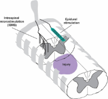
          Neuroprosthetic approaches have tremendous potential for the treatment of injuries to the brain and spinal cord by inducing appropriate neural activity in otherwise disordered circuits. Substantial work has demonstrated that stimulation applied to both the central and peripheral nervous system leads to immediate and in some cases sustained benefits after injury. Here we focus on cervical intraspinal microstimulation (ISMS) as a promising method of activating the spinal cord distal to an injury site, either to directly produce movements or more intriguingly to improve subsequent volitional control of the paretic extremities. Incomplete injuries to the spinal cord are the most commonly observed in human patients, and these injuries spare neural tissue bypassing the lesion that could be influenced by neural devices to promote recovery of function. In fact, recent results have demonstrated that therapeutic ISMS leads to modest but sustained improvements in forelimb function after an incomplete spinal cord injury (SCI). This therapeutic spinal stimulation may promote long-term recovery of function by providing the necessary electrical activity needed for neuron survival, axon growth, and synaptic stability.

          Related collections

          Most cited references61

          • Record: found
          • Abstract: found
          • Article: not found

          Glial regulation of the cerebral microvasculature.

          The brain is a heterogeneous organ with regionally varied and constantly changing energetic needs. Blood vessels in the brain are equipped with control mechanisms that match oxygen and glucose delivery through blood flow with the local metabolic demands that are imposed by neural activity. However, the cellular bases of this mechanism have remained elusive. A major advance has been the demonstration that astrocytes, cells with extensive contacts with both synapses and cerebral blood vessels, participate in the increases in flow evoked by synaptic activity. Their organization in nonoverlapping spatial domains indicates that they are uniquely positioned to shape the spatial distribution of the vascular responses that are evoked by neural activity. Astrocytic calcium is an important determinant of microvascular function and may regulate flow independently of synaptic activity. The involvement of astrocytes in neurovascular coupling has broad implications for the interpretation of functional imaging signals and for the understanding of brain diseases that are associated with neurovascular dysfunction.
            Bookmark
            • Record: found
            • Abstract: found
            • Article: not found

            cAMP and Schwann cells promote axonal growth and functional recovery after spinal cord injury.

            Central neurons regenerate axons if a permissive environment is provided; after spinal cord injury, however, inhibitory molecules are present that make the local environment nonpermissive. A promising new strategy for inducing neurons to overcome inhibitory signals is to activate cAMP signaling. Here we show that cAMP levels fall in the rostral spinal cord, sensorimotor cortex and brainstem after spinal cord contusion. Inhibition of cAMP hydrolysis by the phosphodiesterase IV inhibitor rolipram prevents this decrease and when combined with Schwann cell grafts promotes significant supraspinal and proprioceptive axon sparing and myelination. Furthermore, combining rolipram with an injection of db-cAMP near the graft not only prevents the drop in cAMP levels but increases them above those in uninjured controls. This further enhances axonal sparing and myelination, promotes growth of serotonergic fibers into and beyond grafts, and significantly improves locomotion. These findings show that cAMP levels are key for protection, growth and myelination of injured CNS axons in vivo and recovery of function.
              Bookmark
              • Record: found
              • Abstract: found
              • Article: not found

              Spinal axon regeneration induced by elevation of cyclic AMP.

              Myelin inhibitors, including MAG, are major impediments to CNS regeneration. However, CNS axons of DRGs regenerate if the peripheral branch of these neurons is lesioned first. We show that 1 day post-peripheral-lesion, DRG-cAMP levels triple and MAG/myelin no longer inhibit growth, an effect that is PKA dependent. By 1 week post-lesion, DRG-cAMP returns to control, but growth on MAG/myelin improves and is now PKA independent. Inhibiting PKA in vivo blocks the post-lesion growth on MAG/myelin at 1 day and attenuates it at 1 week. Alone, injection of db-cAMP into the DRG mimics completely a conditioning lesion as DRGs grow on MAG/myelin, initially, in a PKA-dependent manner that becomes PKA independent. Importantly, DRG injection of db-cAMP results in extensive regeneration of dorsal column axons lesioned 1 week later. These results may be relevant to developing therapies for spinal cord injury.
                Bookmark

                Author and article information

                Journal
                Front Neurosci
                Front Neurosci
                Front. Neurosci.
                Frontiers in Neuroscience
                Frontiers Media S.A.
                1662-4548
                1662-453X
                05 January 2014
                27 February 2014
                2014
                : 8
                : 21
                Affiliations
                [1] 1Department of Rehabilitation Medicine, University of Washington Seattle, WA, USA
                [2] 2Department of Neurological Surgery, University of Washington Seattle, WA, USA
                [3] 3Center for Sensorimotor Neural Engineering, University of Washington Seattle, WA, USA
                [4] 4Graduate Program in Neurobiology and Behavior, University of Washington Seattle, WA, USA
                [5] 5Department of Physiology and Biophysics, University of Washington Seattle, WA, USA
                Author notes

                Edited by: Mesut Sahin, New Jersey Institute of Technology, USA

                Reviewed by: Reinhold Scherer, Graz University of Technology, Austria; Andrew Jackson, Newcastle University, UK

                *Correspondence: Chet T. Moritz, Departments of Rehabilitation Medicine and Physiology and Biophysics, School of Medicine, UW Medical Center, University of Washington, 1959 N.E. Pacific Street, PO Box 356490, Seattle, WA 98195, USA e-mail: ctmoritz@ 123456uw.edu

                This article was submitted to Neuroprosthetics, a section of the journal Frontiers in Neuroscience.

                Article
                10.3389/fnins.2014.00021
                3936503
                24578680
                298b7bba-3ab0-4137-bb1b-1441428381a8
                Copyright © 2014 Mondello, Kasten, Horner and Moritz.

                This is an open-access article distributed under the terms of the Creative Commons Attribution License (CC BY). The use, distribution or reproduction in other forums is permitted, provided the original author(s) or licensor are credited and that the original publication in this journal is cited, in accordance with accepted academic practice. No use, distribution or reproduction is permitted which does not comply with these terms.

                History
                : 02 December 2013
                : 24 January 2014
                Page count
                Figures: 2, Tables: 0, Equations: 0, References: 79, Pages: 7, Words: 6010
                Categories
                Neuroscience
                Perspective Article

                Neurosciences
                neuroprosthesis,isms,spinal cord injury,regenerative stimulation,electrical activity
                Neurosciences
                neuroprosthesis, isms, spinal cord injury, regenerative stimulation, electrical activity

                Comments

                Comment on this article