301
views
0
recommends
+1 Recommend
0 collections
    0
    shares
      • Record: found
      • Abstract: found
      • Article: found
      Is Open Access

      Architecture of the RNA polymerase II–TFIIF complex revealed by cross-linking and mass spectrometry

      research-article

      Read this article at

      Bookmark
          There is no author summary for this article yet. Authors can add summaries to their articles on ScienceOpen to make them more accessible to a non-specialist audience.

          Abstract

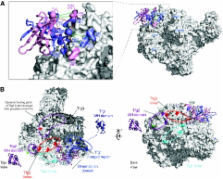
          Higher-order multi-protein complexes such as RNA polymerase II (Pol II) complexes with transcription initiation factors are often not amenable to X-ray structure determination. Here, we show that protein cross-linking coupled to mass spectrometry (MS) has now sufficiently advanced as a tool to extend the Pol II structure to a 15-subunit, 670 kDa complex of Pol II with the initiation factor TFIIF at peptide resolution. The N-terminal regions of TFIIF subunits Tfg1 and Tfg2 form a dimerization domain that binds the Pol II lobe on the Rpb2 side of the active centre cleft near downstream DNA. The C-terminal winged helix (WH) domains of Tfg1 and Tfg2 are mobile, but the Tfg2 WH domain can reside at the Pol II protrusion near the predicted path of upstream DNA in the initiation complex. The linkers between the dimerization domain and the WH domains in Tfg1 and Tfg2 are located to the jaws and protrusion, respectively. The results suggest how TFIIF suppresses non-specific DNA binding and how it helps to recruit promoter DNA and to set the transcription start site. This work establishes cross-linking/MS as an integrated structure analysis tool for large multi-protein complexes.

          Related collections

          Most cited references56

          • Record: found
          • Abstract: found
          • Article: not found

          New yeast-Escherichia coli shuttle vectors constructed with in vitro mutagenized yeast genes lacking six-base pair restriction sites.

          We describe the production of new alleles of the LEU2, URA3 and TRP1 genes of Saccharomyces cerevisiae by in vitro mutagenesis. Each new allele, which lacks restriction enzyme recognition sequences found in the pUC19 multicloning site, was used to construct a unique series of yeast-Escherichia coli shuttle vectors derived from the plasmid pUC19. For each gene a 2 mu vector (YEplac), an ARS1 CEN4 vector (YCplac) and an integrative vector (YIplac) was constructed. The features of these vectors include (i) small size; (ii) unique recognition site for each restriction enzyme found in the pUC19 multicloning site; (iii) screening for plasmids containing inserts by color assay; (iv) high plasmid yield; (v) efficient transformation of S. cerevisiae. These vectors should allow greater flexibility with regard to DNA restriction fragment manipulation and subcloning.
            Bookmark
            • Record: found
            • Abstract: found
            • Article: not found

            Structural basis of transcription: RNA polymerase II at 2.8 angstrom resolution.

            Structures of a 10-subunit yeast RNA polymerase II have been derived from two crystal forms at 2.8 and 3.1 angstrom resolution. Comparison of the structures reveals a division of the polymerase into four mobile modules, including a clamp, shown previously to swing over the active center. In the 2.8 angstrom structure, the clamp is in an open state, allowing entry of straight promoter DNA for the initiation of transcription. Three loops extending from the clamp may play roles in RNA unwinding and DNA rewinding during transcription. A 2.8 angstrom difference Fourier map reveals two metal ions at the active site, one persistently bound and the other possibly exchangeable during RNA synthesis. The results also provide evidence for RNA exit in the vicinity of the carboxyl-terminal repeat domain, coupling synthesis to RNA processing by enzymes bound to this domain.
              Bookmark
              • Record: found
              • Abstract: found
              • Article: not found

              Implications for kinetochore-microtubule attachment from the structure of an engineered Ndc80 complex.

              Kinetochores are proteinaceous assemblies that mediate the interaction of chromosomes with the mitotic spindle. The 180 kDa Ndc80 complex is a direct point of contact between kinetochores and microtubules. Its four subunits contain coiled coils and form an elongated rod structure with functional globular domains at either end. We crystallized an engineered "bonsai" Ndc80 complex containing a shortened rod domain but retaining the globular domains required for kinetochore localization and microtubule binding. The structure reveals a microtubule-binding interface containing a pair of tightly interacting calponin-homology (CH) domains with a previously unknown arrangement. The interaction with microtubules is cooperative and predominantly electrostatic. It involves positive charges in the CH domains and in the N-terminal tail of the Ndc80 subunit and negative charges in tubulin C-terminal tails and is regulated by the Aurora B kinase. We discuss our results with reference to current models of kinetochore-microtubule attachment and centromere organization.
                Bookmark

                Author and article information

                Journal
                EMBO J
                The EMBO Journal
                Nature Publishing Group
                0261-4189
                1460-2075
                17 February 2010
                21 January 2010
                21 January 2010
                : 29
                : 4
                : 717-726
                Affiliations
                [1 ]Wellcome Trust Centre for Cell Biology, Institute of Cell Biology, The University of Edinburgh, Edinburgh, UK
                [2 ]Department of Biochemistry, Gene Center and Center for Integrated Protein Science Munich (CIPSM), Ludwig-Maximilians-Universität München, Munich, Germany
                [3 ]Centre for Systems Biology, The University of Edinburgh, Edinburgh, UK
                [4 ]Department of Structural Biology and Chemistry, Institut Pasteur, Paris, Cedex, France
                Author notes
                [a ]Wellcome Trust Centre for Cell Biology, Institute of Cell Biology, The University of Edinburgh, King's Bldgs, Edinburgh EH9 3JR, UK. Tel.: +44 131 651 7056; Fax: +44 131 650 5379; E-mail: Juri.Rappsilber@ 123456ed.ac.uk
                Article
                emboj2009401
                10.1038/emboj.2009.401
                2810376
                20094031
                2b18cc75-9b8d-48c8-b211-55a2d63f8a7a
                Copyright © 2010, European Molecular Biology Organization

                This is an open-access article distributed under the terms of the Creative Commons Attribution License, which permits distribution, and reproduction in any medium, provided the original author and source are credited. This license does not permit commercial exploitation or the creation of derivative works without specific permission.

                History
                : 13 October 2009
                : 10 December 2009
                Page count
                Pages: 10
                Categories
                Article

                Molecular biology
                higher-order protein complex,integrated structure analysis,mass spectrometry,transcription and its regulation,multi-dimensional structure and dynamics of biological macromolecules

                Comments

                Comment on this article