17
views
0
recommends
+1 Recommend
0 collections
    0
    shares
      • Record: found
      • Abstract: found
      • Article: found
      Is Open Access

      Automatic Segmentation of Anatomical Structures from CT Scans of Thorax for RTP

      research-article

      Read this article at

      Bookmark
          There is no author summary for this article yet. Authors can add summaries to their articles on ScienceOpen to make them more accessible to a non-specialist audience.

          Abstract

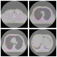
          Modern radiotherapy techniques are vulnerable to delineation inaccuracies owing to the steep dose gradient around the target. In this aspect, accurate contouring comprises an indispensable part of optimal radiation treatment planning (RTP). We suggest a fully automated method to segment the lungs, trachea/main bronchi, and spinal canal accurately from computed tomography (CT) scans of patients with lung cancer to use for RTP. For this purpose, we developed a new algorithm for inclusion of excluded pathological areas into the segmented lungs and a modified version of the fuzzy segmentation by morphological reconstruction for spinal canal segmentation and implemented some image processing algorithms along with them. To assess the accuracy, we performed two comparisons between the automatically obtained results and the results obtained manually by an expert. The average volume overlap ratio values range between 94.30 ± 3.93% and 99.11 ± 0.26% on the two different datasets. We obtained the average symmetric surface distance values between the ranges of 0.28 ± 0.21 mm and 0.89 ± 0.32 mm by using the same datasets. Our method provides favorable results in the segmentation of CT scans of patients with lung cancer and can avoid heavy computational load and might offer expedited segmentation that can be used in RTP.

          Related collections

          Most cited references33

          • Record: found
          • Abstract: found
          • Article: not found

          Comparison and evaluation of methods for liver segmentation from CT datasets.

          This paper presents a comparison study between 10 automatic and six interactive methods for liver segmentation from contrast-enhanced CT images. It is based on results from the "MICCAI 2007 Grand Challenge" workshop, where 16 teams evaluated their algorithms on a common database. A collection of 20 clinical images with reference segmentations was provided to train and tune algorithms in advance. Participants were also allowed to use additional proprietary training data for that purpose. All teams then had to apply their methods to 10 test datasets and submit the obtained results. Employed algorithms include statistical shape models, atlas registration, level-sets, graph-cuts and rule-based systems. All results were compared to reference segmentations five error measures that highlight different aspects of segmentation accuracy. All measures were combined according to a specific scoring system relating the obtained values to human expert variability. In general, interactive methods reached higher average scores than automatic approaches and featured a better consistency of segmentation quality. However, the best automatic methods (mainly based on statistical shape models with some additional free deformation) could compete well on the majority of test images. The study provides an insight in performance of different segmentation approaches under real-world conditions and highlights achievements and limitations of current image analysis techniques.
            Bookmark
            • Record: found
            • Abstract: found
            • Article: not found

            Active shape model segmentation with optimal features.

            An active shape model segmentation scheme is presented that is steered by optimal local features, contrary to normalized first order derivative profiles, as in the original formulation [Cootes and Taylor, 1995, 1999, and 2001]. A nonlinear kNN-classifier is used, instead of the linear Mahalanobis distance, to find optimal displacements for landmarks. For each of the landmarks that describe the shape, at each resolution level taken into account during the segmentation optimization procedure, a distinct set of optimal features is determined. The selection of features is automatic, using the training images and sequential feature forward and backward selection. The new approach is tested on synthetic data and in four medical segmentation tasks: segmenting the right and left lung fields in a database of 230 chest radiographs, and segmenting the cerebellum and corpus callosum in a database of 90 slices from MRI brain images. In all cases, the new method produces significantly better results in terms of an overlap error measure (p < 0.001 using a paired T-test) than the original active shape model scheme.
              Bookmark
              • Record: found
              • Abstract: found
              • Article: not found

              Automatic lung segmentation from thoracic computed tomography scans using a hybrid approach with error detection.

              Lung segmentation is a prerequisite for automated analysis of chest CT scans. Conventional lung segmentation methods rely on large attenuation differences between lung parenchyma and surrounding tissue. These methods fail in scans where dense abnormalities are present, which often occurs in clinical data. Some methods to handle these situations have been proposed, but they are too time consuming or too specialized to be used in clinical practice. In this article, a new hybrid lung segmentation method is presented that automatically detects failures of a conventional algorithm and, when needed, resorts to a more complex algorithm, which is expected to produce better results in abnormal cases. In a large quantitative evaluation on a database of 150 scans from different sources, the hybrid method is shown to perform substantially better than a conventional approach at a relatively low increase in computational cost.
                Bookmark

                Author and article information

                Journal
                Comput Math Methods Med
                Comput Math Methods Med
                CMMM
                Computational and Mathematical Methods in Medicine
                Hindawi Publishing Corporation
                1748-670X
                1748-6718
                2014
                18 December 2014
                : 2014
                : 472890
                Affiliations
                1Electrical and Electronics Engineering Department, Faculty of Engineering, Ankara University, Gölbaşı, 06830 Ankara, Turkey
                2Radiation Oncology Department, Gülhane Military Medical Academy, Etlik, 06018 Ankara, Turkey
                Author notes
                *Emin Emrah Özsavaş: emrah605@ 123456yahoo.com

                Academic Editor: Chuangyin Dang

                Article
                10.1155/2014/472890
                4281476
                2d272a1e-fcf3-43a2-8f97-138ba56014ba
                Copyright © 2014 Emin Emrah Özsavaş et al.

                This is an open access article distributed under the Creative Commons Attribution License, which permits unrestricted use, distribution, and reproduction in any medium, provided the original work is properly cited.

                History
                : 30 July 2014
                : 26 November 2014
                : 1 December 2014
                Categories
                Research Article

                Applied mathematics
                Applied mathematics

                Comments

                Comment on this article