30
views
0
recommends
+1 Recommend
0 collections
    0
    shares
      • Record: found
      • Abstract: found
      • Article: found
      Is Open Access

      Control of Vascular Smooth Muscle Cell Growth by Connexin 43

      research-article

      Read this article at

      Bookmark
          There is no author summary for this article yet. Authors can add summaries to their articles on ScienceOpen to make them more accessible to a non-specialist audience.

          Abstract

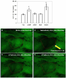
          Connexin 43 (Cx43), the principal gap junction protein in vascular smooth muscle cells (VSMCs), regulates movement of ions and other signaling molecules through gap junction intercellular communication (GJIC) and plays important roles in maintaining normal vessel function; however, many of the signaling mechanisms controlling Cx43 in VSMCs are not clearly described. The goal of this study was to investigate mechanisms of Cx43 regulation with respect to VSMC proliferation. Treatment of rat primary VSMCs with the cAMP analog 8Br-cAMP, the soluble guanylate cyclase (sGC) stimulator BAY 41-2272 (BAY), or the Cx inducer diallyl disulfide (DADS) significantly reduced proliferation after 72 h compared with vehicle controls. Bromodeoxyuridine uptake revealed reduction ( p < 0.05) in DNA synthesis after 6 h and flow cytometry showed reduced (40%) S-phase cell numbers after 16 h in DADS-treated cells compared with vehicle controls. Cx43 expression significantly increased after 270 min treatment with 8Br-cAMP, 8Br-cGMP, BAY or DADS. Inhibition of PKA, PKG or PKC reversed 8Br-cAMP-stimulated increases in Cx43 expression, whereas only PKG or PKC inhibition reversed 8Br-cGMP- and BAY-stimulated increases in total Cx43. Interestingly, stimulation of Cx43 expression by DADS was not dependent on PKA, PKG or PKC. Using fluorescence recovery after photobleaching, only 8Br-cAMP or DADS increased GJIC with 8Br-cAMP mediated by PKC and DADS mediated by PKG. Further, DADS significantly increased phosphorylation at MAPK-sensitive Serine (Ser)255 and Ser279, the cell cycle regulatory kinase-sensitive Ser262 and PKC-sensitive Ser368 after 30 min while 8Br-cAMP significantly increased phosphorylation only at Ser279 compared with controls. This study demonstrates that 8Br-cAMP- and DADS-enhanced GJIC rather than Cx43 expression and/or phosphorylation plays important roles in the regulation of VSMC proliferation and provides new insights into the growth-regulatory capacities of Cx43 in VSM.

          Related collections

          Most cited references76

          • Record: found
          • Abstract: not found
          • Article: not found

          The gap junction communication channel.

            Bookmark
            • Record: found
            • Abstract: found
            • Article: not found

            Gap junctions: structure and function (Review).

            Gap junctions are plasma membrane spatial microdomains constructed of assemblies of channel proteins called connexins in vertebrates and innexins in invertebrates. The channels provide direct intercellular communication pathways allowing rapid exchange of ions and metabolites up to approximately 1 kD in size. Approximately 20 connexins are identified in the human or mouse genome, and orthologues are increasingly characterized in other vertebrates. Most cell types express multiple connexin isoforms, making likely the construction of a spectrum of heteromeric hemichannels and heterotypic gap junctions that could provide a structural basis for the charge and size selectivity of these intercellular channels. The precise nature of the potential signalling information traversing junctions in physiologically defined situations remains elusive, but extensive progress has been made in elucidating how connexins are assembled into gap junctions. Also, participation of gap junction hemichannels in the propagation of calcium waves via an extracellular purinergic pathway is emerging. Connexin mutations have been identified in a number of genetically inherited channel communication-opathies. These are detected in connexin 32 in Charcot Marie Tooth-X linked disease, in connexins 26 and 30 in deafness and skin diseases, and in connexins 46 and 50 in hereditary cataracts. Biochemical approaches indicate that many of the mutated connexins are mistargeted to gap junctions and/or fail to oligomerize correctly into hemichannels. Genetic ablation approaches are helping to map out a connexin code and point to specific connexins being required for cell growth and differentiation as well as underwriting basic intercellular communication.
              Bookmark
              • Record: found
              • Abstract: found
              • Article: not found

              The effects of connexin phosphorylation on gap junctional communication.

              Gap junctions are specialized membrane domains composed of collections of channels that directly connect neighboring cells providing for the cell-to-cell diffusion of small molecules, including ions, amino acids, nucleotides, and second messengers. Vertebrate gap junctions are composed of proteins encoded by the "connexin" gene family. In most cases examined, connexins are modified post-translationally by phosphorylation. Phosphorylation has been implicated in the regulation of gap junctional communication at several stages of the connexin "lifecycle", such as the trafficking, assembly/disassembly, degradation, as well as, the gating of gap junction channels. Since connexin43 (Cx43) is widely expressed in tissues and cell lines, we understand the most about how it is regulated, and thus, connexin43 phosphorylation is a major focus of this review. Recent reports utilizing new methodologies combined with the latest genome information have shown that activation of several kinases including protein kinase A, protein kinase C, p34(cdc2)/cyclin B kinase, casein kinase 1, mitogen-activated protein (MAP) kinase and pp60(src) kinase can lead to phosphorylation at 12 of the 21 serine and two of the six tyrosine residues in the C-terminal region of connexin43. In several cases, use of site-directed mutants of these sites have shown that these specific phosphorylation events can be linked to changes in gap junctional communication.
                Bookmark

                Author and article information

                Journal
                Front Physiol
                Front Physiol
                Front. Physio.
                Frontiers in Physiology
                Frontiers Research Foundation
                1664-042X
                21 June 2012
                2012
                : 3
                : 220
                Affiliations
                [1] 1simpleDepartment of Physiology, Brody School of Medicine, East Carolina University Greenville, NC, USA
                [2] 2simpleDepartment of Cardiovascular Sciences, Brody School of Medicine, East Carolina University Greenville, NC, USA
                Author notes

                Edited by: Timothy R. Nurkiewicz, West Virginia University, USA

                Reviewed by: Timothy R. Nurkiewicz, West Virginia University, USA; Daniel Joseph Conklin, University of Louisville, USA

                *Correspondence: David A. Tulis, Department of Physiology, Brody School of Medicine, East Carolina University, 6E-108, 600 Moye Boulevard, Greenville, NC 27834, USA. e-mail: tulisd@ 123456ecu.edu

                This article was submitted to Frontiers in Vascular Physiology, a specialty of Frontiers in Physiology.

                Article
                10.3389/fphys.2012.00220
                3380337
                22737133
                2dd30163-f6b3-464f-a551-9b41f804613c
                Copyright © 2012 Joshi, Martin, Shaver, Madamanchi, Muller-Borer and Tulis.

                This is an open-access article distributed under the terms of the Creative Commons Attribution Non Commercial License, which permits non-commercial use, distribution, and reproduction in other forums, provided the original authors and source are credited.

                History
                : 04 April 2012
                : 01 June 2012
                Page count
                Figures: 8, Tables: 1, Equations: 0, References: 77, Pages: 13, Words: 9573
                Categories
                Physiology
                Original Research

                Anatomy & Physiology
                vascular smooth muscle cells,cx43,cgmp,camp,protein kinases
                Anatomy & Physiology
                vascular smooth muscle cells, cx43, cgmp, camp, protein kinases

                Comments

                Comment on this article