3
views
0
recommends
+1 Recommend
0 collections
    0
    shares
      • Record: found
      • Abstract: found
      • Article: not found

      Diagnosis of breast cancer based on modern mammography using hybrid transfer learning

      research-article

      Read this article at

      ScienceOpenPublisherPMC
      Bookmark
          There is no author summary for this article yet. Authors can add summaries to their articles on ScienceOpen to make them more accessible to a non-specialist audience.

          Abstract

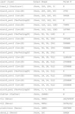
          Breast cancer is a common cancer in women. Early detection of breast cancer in particular and cancer, in general, can considerably increase the survival rate of women, and it can be much more effective. This paper mainly focuses on the transfer learning process to detect breast cancer. Modified VGG (MVGG) is proposed and implemented on datasets of 2D and 3D images of mammograms. Experimental results showed that the proposed hybrid transfer learning model (a fusion of MVGG and ImageNet) provides an accuracy of 94.3%. On the other hand, only the proposed MVGG architecture provides an accuracy of 89.8%. So, it is precisely stated that the proposed hybrid pre-trained network outperforms other compared Convolutional Neural Networks. The proposed architecture can be considered as an effective tool for radiologists to decrease the false negative and false positive rates. Therefore, the efficiency of mammography analysis will be improved.

          Related collections

          Most cited references27

          • Record: found
          • Abstract: found
          • Article: not found

          Comparison of the performance of screening mammography, physical examination, and breast US and evaluation of factors that influence them: an analysis of 27,825 patient evaluations.

          To (a) determine the performance of screening mammography, ultrasonography (US), and physical examination (PE); (b) analyze the influence of age, hormonal status, and breast density; (c) compare the size and stage of tumors detected with each modality; and (d) determine which modality or combination of modalities optimize cancer detection. A total of 11,130 asymptomatic women underwent 27,825 screening sessions, (mammography and subsequent PE). Women with dense breasts subsequently underwent screening US. Abnormalities were deemed positive if biopsy findings revealed malignancy and negative if findings from biopsy or all screening examinations were negative. In 221 women, 246 cancers were found. Sensitivity, specificity, negative and positive predictive values, and accuracy of mammography were 77.6%, 98.8%, 99.8%, 35.8%, and 98.6%, respectively; those of PE, 27.6%, 99.4%, 99.4%, 28.9%, and 98.8%, respectively; and those of US, 75.3%, 96.8%, 99.7%, 20.5%, and 96.6%, respectively. Screening breast US increased the number of women diagnosed with nonpalpable invasive cancers by 42% (30 of 71). Mammographic sensitivity declined significantly with increasing breast density (P <.01) (48% for the densest breasts) and in younger women with dense breasts (P =.02); the effects were independent. Mammography and US together had significantly higher sensitivity (97%) than did mammography and PE together (74%) (P <.001). Tumors detected at mammography and/or US were significantly smaller (P =.01) and of lower stage (P =.01) than those detected at PE. Mammographic sensitivity for breast cancer declines significantly with increasing breast density and is independently higher in older women with dense breasts. Addition of screening US significantly increases detection of small cancers and depicts significantly more cancers and at smaller size and lower stage than does PE, which detects independently extremely few cancers. Hormonal status has no significant effect on effectiveness of screening independent of breast density.
            Bookmark
            • Record: found
            • Abstract: found
            • Article: not found

            Lymph Node Metastasis Prediction from Primary Breast Cancer US Images Using Deep Learning.

            Background Deep learning (DL) algorithms are gaining extensive attention for their excellent performance in image recognition tasks. DL models can automatically make a quantitative assessment of complex medical image characteristics and achieve increased accuracy in diagnosis with higher efficiency. Purpose To determine the feasibility of using a DL approach to predict clinically negative axillary lymph node metastasis from US images in patients with primary breast cancer. Materials and Methods A data set of US images in patients with primary breast cancer with clinically negative axillary lymph nodes from Tongji Hospital (974 imaging studies from 2016 to 2018, 756 patients) and an independent test set from Hubei Cancer Hospital (81 imaging studies from 2018 to 2019, 78 patients) were collected. Axillary lymph node status was confirmed with pathologic examination. Three different convolutional neural networks (CNNs) of Inception V3, Inception-ResNet V2, and ResNet-101 architectures were trained on 90% of the Tongji Hospital data set and tested on the remaining 10%, as well as on the independent test set. The performance of the models was compared with that of five radiologists. The models' performance was analyzed in terms of accuracy, sensitivity, specificity, receiver operating characteristic curves, areas under the receiver operating characteristic curve (AUCs), and heat maps. Results The best-performing CNN model, Inception V3, achieved an AUC of 0.89 (95% confidence interval [CI]: 0.83, 0.95) in the prediction of the final clinical diagnosis of axillary lymph node metastasis in the independent test set. The model achieved 85% sensitivity (35 of 41 images; 95% CI: 70%, 94%) and 73% specificity (29 of 40 images; 95% CI: 56%, 85%), and the radiologists achieved 73% sensitivity (30 of 41 images; 95% CI: 57%, 85%; P = .17) and 63% specificity (25 of 40 images; 95% CI: 46%, 77%; P = .34). Conclusion Using US images from patients with primary breast cancer, deep learning models can effectively predict clinically negative axillary lymph node metastasis. Artificial intelligence may provide an early diagnostic strategy for lymph node metastasis in patients with breast cancer with clinically negative lymph nodes. Published under a CC BY 4.0 license. Online supplemental material is available for this article. See also the editorial by Bae in this issue.
              Bookmark
              • Record: found
              • Abstract: found
              • Article: found
              Is Open Access

              A curated mammography data set for use in computer-aided detection and diagnosis research

              Published research results are difficult to replicate due to the lack of a standard evaluation data set in the area of decision support systems in mammography; most computer-aided diagnosis (CADx) and detection (CADe) algorithms for breast cancer in mammography are evaluated on private data sets or on unspecified subsets of public databases. This causes an inability to directly compare the performance of methods or to replicate prior results. We seek to resolve this substantial challenge by releasing an updated and standardized version of the Digital Database for Screening Mammography (DDSM) for evaluation of future CADx and CADe systems (sometimes referred to generally as CAD) research in mammography. Our data set, the CBIS-DDSM (Curated Breast Imaging Subset of DDSM), includes decompressed images, data selection and curation by trained mammographers, updated mass segmentation and bounding boxes, and pathologic diagnosis for training data, formatted similarly to modern computer vision data sets. The data set contains 753 calcification cases and 891 mass cases, providing a data-set size capable of analyzing decision support systems in mammography.
                Bookmark

                Author and article information

                Contributors
                thanhdnh@ueh.edu.vn
                Journal
                Multidimens Syst Signal Process
                Multidimens Syst Signal Process
                Multidimensional Systems and Signal Processing
                Springer US (New York )
                0923-6082
                1573-0824
                11 January 2021
                : 1-19
                Affiliations
                [1 ]GRID grid.449005.c, School of Computer Science and Engineering, , Lovely Professional University, ; Phagwara, Punjab India
                [2 ]GRID grid.411512.2, ISNI 0000 0001 2223 0518, Institute of Information and Communication Technology, , Bangladesh University of Engineering and Technology, ; Dhaka, 1205 Bangladesh
                [3 ]GRID grid.411685.f, ISNI 0000 0004 0498 1133, Maharaja Agrasen Institute of Technology, ; Delhi, India
                [4 ]GRID grid.444827.9, ISNI 0000 0000 9009 5680, School of Business Information Technology, , University of Economics Ho Chi Minh City, ; Ho Chi Minh City, Vietnam
                Author information
                http://orcid.org/0000-0003-2025-8319
                Article
                756
                10.1007/s11045-020-00756-7
                7798373
                33456204
                2ff95109-f417-4178-9e49-a6b3ada68fd7
                © The Author(s), under exclusive licence to Springer Science+Business Media, LLC part of Springer Nature 2021

                This article is made available via the PMC Open Access Subset for unrestricted research re-use and secondary analysis in any form or by any means with acknowledgement of the original source. These permissions are granted for the duration of the World Health Organization (WHO) declaration of COVID-19 as a global pandemic.

                History
                : 24 May 2020
                : 9 December 2020
                : 19 December 2020
                Categories
                Article

                hybrid transfer learning,medical image segmentation,breast cancer,mammography,3d mammography,convolutional neural networks

                Comments

                Comment on this article