271
views
0
recommends
+1 Recommend
0 collections
    0
    shares
      • Record: found
      • Abstract: found
      • Article: not found

      Optogenetic stimulation of a hippocampal engram activates fear memory recall

      research-article

      Read this article at

      ScienceOpenPublisherPMC
      Bookmark
          There is no author summary for this article yet. Authors can add summaries to their articles on ScienceOpen to make them more accessible to a non-specialist audience.

          Abstract

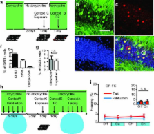
          A specific memory is thought to be encoded by a sparse population of neurons 1, 2 . These neurons can be tagged during learning for subsequent identification 3 and manipulation 4, 5, 6 . Moreover, their ablation or inactivation results in reduced memory expression, suggesting their necessity in mnemonic processes. However, a critical question of sufficiency remains: can one elicit the behavioral output of a specific memory by directly activating a population of neurons that was active during learning? Here we show that optogenetic reactivation of hippocampal neurons activated during fear conditioning is sufficient to induce freezing behavior. We labeled a population of hippocampal dentate gyrus neurons activated during fear learning with channelrhodopsin-2 (ChR2) 7, 8 and later optically reactivated these neurons in a different context. The mice showed increased freezing only upon light stimulation, indicating light-induced fear memory recall. This freezing was not detected in non-fear conditioned mice expressing ChR2 in a similar proportion of cells, nor in fear conditioned mice with cells labeled by EYFP instead of ChR2. Finally, activation of cells labeled in a context not associated with fear did not evoke freezing in mice that were previously fear conditioned in a different context, suggesting that light-induced fear memory recall is context-specific. Together, our findings indicate that activating a sparse but specific ensemble of hippocampal neurons that contribute to a memory engram is sufficient for the recall of that memory. Moreover, our experimental approach offers a general method of mapping cellular populations bearing memory engrams.

          Related collections

          Most cited references28

          • Record: found
          • Abstract: found
          • Article: not found

          Pattern separation in the dentate gyrus and CA3 of the hippocampus.

          Theoretical models have long pointed to the dentate gyrus as a possible source of neuronal pattern separation. In agreement with predictions from these models, we show that minimal changes in the shape of the environment in which rats are exploring can substantially alter correlated activity patterns among place-modulated granule cells in the dentate gyrus. When the environments are made more different, new cell populations are recruited in CA3 but not in the dentate gyrus. These results imply a dual mechanism for pattern separation in which signals from the entorhinal cortex can be decorrelated both by changes in coincidence patterns in the dentate gyrus and by recruitment of nonoverlapping cell assemblies in CA3.
            Bookmark
            • Record: found
            • Abstract: found
            • Article: not found

            Amygdala circuitry mediating reversible and bidirectional control of anxiety.

            Anxiety--a sustained state of heightened apprehension in the absence of immediate threat--becomes severely debilitating in disease states. Anxiety disorders represent the most common of psychiatric diseases (28% lifetime prevalence) and contribute to the aetiology of major depression and substance abuse. Although it has been proposed that the amygdala, a brain region important for emotional processing, has a role in anxiety, the neural mechanisms that control anxiety remain unclear. Here we explore the neural circuits underlying anxiety-related behaviours by using optogenetics with two-photon microscopy, anxiety assays in freely moving mice, and electrophysiology. With the capability of optogenetics to control not only cell types but also specific connections between cells, we observed that temporally precise optogenetic stimulation of basolateral amygdala (BLA) terminals in the central nucleus of the amygdala (CeA)--achieved by viral transduction of the BLA with a codon-optimized channelrhodopsin followed by restricted illumination in the downstream CeA--exerted an acute, reversible anxiolytic effect. Conversely, selective optogenetic inhibition of the same projection with a third-generation halorhodopsin (eNpHR3.0) increased anxiety-related behaviours. Importantly, these effects were not observed with direct optogenetic control of BLA somata, possibly owing to recruitment of antagonistic downstream structures. Together, these results implicate specific BLA-CeA projections as critical circuit elements for acute anxiety control in the mammalian brain, and demonstrate the importance of optogenetically targeting defined projections, beyond simply targeting cell types, in the study of circuit function relevant to neuropsychiatric disease.
              Bookmark
              • Record: found
              • Abstract: found
              • Article: not found

              Young dentate granule cells mediate pattern separation, whereas old granule cells facilitate pattern completion.

              Adult-born granule cells (GCs), a minor population of cells in the hippocampal dentate gyrus, are highly active during the first few weeks after functional integration into the neuronal network, distinguishing them from less active, older adult-born GCs and the major population of dentate GCs generated developmentally. To ascertain whether young and old GCs perform distinct memory functions, we created a transgenic mouse in which output of old GCs was specifically inhibited while leaving a substantial portion of young GCs intact. These mice exhibited enhanced or normal pattern separation between similar contexts, which was reduced following ablation of young GCs. Furthermore, these mutant mice exhibited deficits in rapid pattern completion. Therefore, pattern separation requires adult-born young GCs but not old GCs, and older GCs contribute to the rapid recall by pattern completion. Our data suggest that as adult-born GCs age, their function switches from pattern separation to rapid pattern completion. Copyright © 2012 Elsevier Inc. All rights reserved.
                Bookmark

                Author and article information

                Journal
                0410462
                6011
                Nature
                Nature
                Nature
                0028-0836
                1476-4687
                16 March 2012
                22 March 2012
                19 October 2012
                : 484
                : 7394
                : 381-385
                Affiliations
                [1 ]RIKEN-MIT Center for Neural Circuit Genetics at the Picower Institute for Learning and Memory, Department of Biology and Department of Brain and Cognitive Sciences, Massachusetts Institute of Technology, Cambridge, MA, U.S.A.
                [2 ]Department of Bioengineering and Department of Psychiatry and Behavioral Sciences, Stanford University, Stanford, CA, U.S.A.
                Author notes
                [*]

                These authors contributed equally to this work.

                [** ]Correspondence and requests for materials should be addressed to S.T. ( tonegawa@ 123456mit.edu ) Reprints and permissions information is available at www.nature.com/reprints.
                Article
                NIHMS364287
                10.1038/nature11028
                3331914
                22441246
                31bbcdc3-8285-484f-94f1-da320649abf8

                Users may view, print, copy, download and text and data- mine the content in such documents, for the purposes of academic research, subject always to the full Conditions of use: http://www.nature.com/authors/editorial_policies/license.html#terms

                History
                Funding
                Funded by: National Institute of Mental Health : NIMH
                Award ID: R01 MH078821-17 || MH
                Funded by: National Institute of Mental Health : NIMH
                Award ID: P50 MH058880-10 || MH
                Categories
                Article

                Uncategorized
                Uncategorized

                Comments

                Comment on this article