8
views
0
recommends
+1 Recommend
0 collections
    0
    shares
      • Record: found
      • Abstract: found
      • Article: found
      Is Open Access

      Brainstem pathology in amyotrophic lateral sclerosis and primary lateral sclerosis: A longitudinal neuroimaging study

      research-article
      a , * , a , a , a , b , b , b , c , d , b , a
      NeuroImage : Clinical
      Elsevier
      Motor neuron disease, Amyotrophic lateral sclerosis, Primary lateral sclerosis, Longitudinal study, Brainstem, Medulla, Pons, Mesencephalon, Neuroimaging, AD, axial diffusivity, ALS, amyotrophic lateral sclerosis, ALS T1, ALS cohort at the first timepoint, ALS T2, ALS cohort at the second timepoint, ANCOVA, analysis of covariance, BG, basal ganglia, BRS, brainstem, C9orf72, chromosome 9 open reading frame 72, CN, cranial nerve, CNN, cranial nerve nuclei, CST, corticospinal tract, Cr, creatine‐phosphocreatine, DTI, diffusion tensor imaging, EPI, echo-planar imaging, FA, fractional anisotropy, FDR, false discovery rate, FTD, frontotemporal dementia, FOV, field-of-view, FSL, FMRIB Software Library, FWE, familywise error, GM, grey matter, HARDI, high angular resolution diffusion imaging, HC, healthy control, HSP, hereditary spastic paraplegia, IR-SPGR, Inversion Recovery prepared Spoiled Gradient Recalled echo, LMN, lower motor neuron, M, mean, MB, midbrain, MEM, mixed-effect model, MD, mean diffusivity, MND, motor neuron disease, MNI152, Montreal Neurological Institute 152 standard space, MR, magnetic resonance, MRS, magnetic resonance spectroscopy, Myo, myoinositol, NAA, N-acetylaspartate, NODDI, Neurite orientation dispersion and density imaging, PBA, pseudobulbar affect, PCL, pathological crying and laughing, PLS, primary lateral sclerosis, PMC, primary motor cortex, pTDP-43, phosphorylated 43 kDa TAR DNA-binding protein, QBI, q-ball imaging, RE, repeat expansion, RD, radial diffusivity, SC, spinal cord, SD, standard deviation, SBMA, spinal and bulbar muscular atrophy / Kennedy's disease, SOD1, superoxide dismutase 1, T1, Timepoint 1, T1W, T1-weighted imaging, T2, Timepoint 2, TBSS, tract-based spatial statistics, TE, echo time, TFCE, threshold-free cluster enhancement, TI, inversion time, TIV, total intracranial volume, TR, repetition time, UMN, upper motor neuron, WM, white matter

      Read this article at

      Bookmark
          There is no author summary for this article yet. Authors can add summaries to their articles on ScienceOpen to make them more accessible to a non-specialist audience.

          Highlights

          • Computational neuroimaging captures focal brainstem pathology in motor neuron diseases in contrast to both healthy- and disease controls.

          • ALS patients exhibit progressive medulla oblongata, pontine and mesencephalic volume loss over time.

          • Brainstem atrophy in ALS and PLS is dominated by medulla oblongata volume reductions.

          • Vertex analyses of ALS patients reveal flattening of the medullary pyramids bilaterally.

          • Morphometric analyses in ALS detect density reductions in the mesencephalic crura consistent with corticospinal tract degeneration.

          Abstract

          Background

          Brainstem pathology is a hallmark feature of ALS, yet most imaging studies focus on cortical grey matter alterations and internal capsule white matter pathology. Brainstem imaging in ALS provides a unique opportunity to appraise descending motor tract degeneration and bulbar lower motor neuron involvement.

          Methods

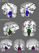
          A prospective longitudinal imaging study has been undertaken with 100 patients with ALS, 33 patients with PLS, 30 patients with FTD and 100 healthy controls. Volumetric, vertex and morphometric analyses were conducted correcting for demographic factors to characterise disease-specific patterns of brainstem pathology. Using a Bayesian segmentation algorithm, the brainstem was segmented into the medulla, pons and mesencephalon to measure regional volume reductions, shape analyses were performed to ascertain the atrophy profile of each study group and region-of-interest morphometry was used to evaluate focal density alterations.

          Results

          ALS and PLS patients exhibit considerable brainstem atrophy compared to both disease- and healthy controls. Volume reductions in ALS and PLS are dominated by medulla oblongata pathology, but pontine atrophy can also be detected. In ALS, vertex analyses confirm the flattening of the medullary pyramids bilaterally in comparison to healthy controls and widespread pontine shape deformations in contrast to PLS. The ALS cohort exhibit bilateral density reductions in the mesencephalic crura in contrast to healthy controls, central pontine atrophy compared to disease controls, peri-aqueduct mesencephalic and posterior pontine changes in comparison to PLS patients.

          Conclus

          ions: Computational brainstem imaging captures the degeneration of both white and grey matter components in ALS. Our longitudinal data indicate progressive brainstem atrophy over time, underlining the biomarker potential of quantitative brainstem measures in ALS. At a time when a multitude of clinical trials are underway worldwide, there is an unprecedented need for accurate biomarkers to monitor disease progression and detect response to therapy. Brainstem imaging is a promising addition to candidate biomarkers of ALS and PLS.

          Related collections

          Most cited references71

          • Record: found
          • Abstract: found
          • Article: not found

          Corpus callosum involvement is a consistent feature of amyotrophic lateral sclerosis.

          While the hallmark of amyotrophic lateral sclerosis (ALS) is corticospinal tract in combination with lower motor neuron degeneration, the clinical involvement of both compartments is characteristically variable and the site of onset debated. We sought to establish whether there is a consistent signature of cerebral white matter abnormalities in heterogeneous ALS cases. In this observational study, diffusion tensor imaging was applied in a whole-brain analysis of 24 heterogeneous patients with ALS and well-matched healthy controls. Tract-based spatial statistics were used, with optimized voxel-based morphometry of T1 images to determine any associated gray matter involvement. A consistent reduction in fractional anisotropy was demonstrated in the corpus callosum of the ALS group, extending rostrally and bilaterally to the region of the primary motor cortices, independent of the degree of clinical upper motor neuron involvement. Matched regional radial diffusivity increase supported the concept of anterograde degeneration of callosal fibers observed pathologically. Gray matter reductions were observed bilaterally in primary motor and supplementary motor regions, and also in the anterior cingulate and temporal lobe regions. A post hoc group comparison model incorporating significant values for fractional anisotropy, radial diffusivity, and gray matter was 92% sensitive, 88% specific, with an accuracy of 90%. Callosal involvement is a consistent feature of ALS, independent of clinical upper motor neuron involvement, and may reflect independent bilateral cortical involvement or interhemispheric spread of pathology. The predominantly rostral corticospinal tract involvement further supports the concept of independent cortical degeneration even in those patients with ALS with predominantly lower motor neuron involvement clinically.
            Bookmark
            • Record: found
            • Abstract: found
            • Article: not found
            Is Open Access

            Bayesian segmentation of brainstem structures in MRI.

            In this paper we present a method to segment four brainstem structures (midbrain, pons, medulla oblongata and superior cerebellar peduncle) from 3D brain MRI scans. The segmentation method relies on a probabilistic atlas of the brainstem and its neighboring brain structures. To build the atlas, we combined a dataset of 39 scans with already existing manual delineations of the whole brainstem and a dataset of 10 scans in which the brainstem structures were manually labeled with a protocol that was specifically designed for this study. The resulting atlas can be used in a Bayesian framework to segment the brainstem structures in novel scans. Thanks to the generative nature of the scheme, the segmentation method is robust to changes in MRI contrast or acquisition hardware. Using cross validation, we show that the algorithm can segment the structures in previously unseen T1 and FLAIR scans with great accuracy (mean error under 1mm) and robustness (no failures in 383 scans including 168 AD cases). We also indirectly evaluate the algorithm with a experiment in which we study the atrophy of the brainstem in aging. The results show that, when used simultaneously, the volumes of the midbrain, pons and medulla are significantly more predictive of age than the volume of the entire brainstem, estimated as their sum. The results also demonstrate that the method can detect atrophy patterns in the brainstem structures that have been previously described in the literature. Finally, we demonstrate that the proposed algorithm is able to detect differential effects of AD on the brainstem structures. The method will be implemented as part of the popular neuroimaging package FreeSurfer.
              Bookmark
              • Record: found
              • Abstract: found
              • Article: not found

              A large-scale multicentre cerebral diffusion tensor imaging study in amyotrophic lateral sclerosis.

              Damage to the cerebral tissue structural connectivity associated with amyotrophic lateral sclerosis (ALS), which extends beyond the motor pathways, can be visualised by diffusion tensor imaging (DTI). The effective translation of DTI metrics as biomarker requires its application across multiple MRI scanners and patient cohorts. A multicentre study was undertaken to assess structural connectivity in ALS within a large sample size.
                Bookmark

                Author and article information

                Contributors
                Journal
                Neuroimage Clin
                Neuroimage Clin
                NeuroImage : Clinical
                Elsevier
                2213-1582
                24 October 2019
                2019
                24 October 2019
                : 24
                : 102054
                Affiliations
                [a ]Computational Neuroimaging Group, Biomedical Sciences Institute, Trinity College Dublin, 152-160 Pearse Street, Dublin 2, Ireland
                [b ]Complex Trait Genomics Laboratory, Smurfit Institute of Genetics, Trinity College Dublin, 152-160 Pearse Street, Dublin 2, Ireland
                [c ]Department of Neurology, St James's Hospital, James's St, Ushers, Dublin 8 D08 NHY1, Ireland
                [d ]Department of Neurology, Western Health & Social Care Trust, Belfast, UK
                Author notes
                [* ]Corresponding author. bedep@ 123456tcd.ie
                Article
                S2213-1582(19)30401-2 102054
                10.1016/j.nicl.2019.102054
                6849418
                31711033
                32f1630c-3590-4050-bd83-62e15ddcff89
                © 2019 The Author(s)

                This is an open access article under the CC BY-NC-ND license (http://creativecommons.org/licenses/by-nc-nd/4.0/).

                History
                : 25 September 2019
                : 10 October 2019
                : 21 October 2019
                Categories
                Regular Article

                motor neuron disease,amyotrophic lateral sclerosis,primary lateral sclerosis,longitudinal study,brainstem,medulla,pons,mesencephalon,neuroimaging,ad, axial diffusivity,als, amyotrophic lateral sclerosis,als t1, als cohort at the first timepoint,als t2, als cohort at the second timepoint,ancova, analysis of covariance,bg, basal ganglia,brs, brainstem,c9orf72, chromosome 9 open reading frame 72,cn, cranial nerve,cnn, cranial nerve nuclei,cst, corticospinal tract,cr, creatine‐phosphocreatine,dti, diffusion tensor imaging,epi, echo-planar imaging,fa, fractional anisotropy,fdr, false discovery rate,ftd, frontotemporal dementia,fov, field-of-view,fsl, fmrib software library,fwe, familywise error,gm, grey matter,hardi, high angular resolution diffusion imaging,hc, healthy control,hsp, hereditary spastic paraplegia,ir-spgr, inversion recovery prepared spoiled gradient recalled echo,lmn, lower motor neuron,m, mean,mb, midbrain,mem, mixed-effect model,md, mean diffusivity,mnd, motor neuron disease,mni152, montreal neurological institute 152 standard space,mr, magnetic resonance,mrs, magnetic resonance spectroscopy,myo, myoinositol,naa, n-acetylaspartate,noddi, neurite orientation dispersion and density imaging,pba, pseudobulbar affect,pcl, pathological crying and laughing,pls, primary lateral sclerosis,pmc, primary motor cortex,ptdp-43, phosphorylated 43 kda tar dna-binding protein,qbi, q-ball imaging,re, repeat expansion,rd, radial diffusivity,sc, spinal cord,sd, standard deviation,sbma, spinal and bulbar muscular atrophy / kennedy's disease,sod1, superoxide dismutase 1,t1, timepoint 1,t1w, t1-weighted imaging,t2, timepoint 2,tbss, tract-based spatial statistics,te, echo time,tfce, threshold-free cluster enhancement,ti, inversion time,tiv, total intracranial volume,tr, repetition time,umn, upper motor neuron,wm, white matter
                motor neuron disease, amyotrophic lateral sclerosis, primary lateral sclerosis, longitudinal study, brainstem, medulla, pons, mesencephalon, neuroimaging, ad, axial diffusivity, als, amyotrophic lateral sclerosis, als t1, als cohort at the first timepoint, als t2, als cohort at the second timepoint, ancova, analysis of covariance, bg, basal ganglia, brs, brainstem, c9orf72, chromosome 9 open reading frame 72, cn, cranial nerve, cnn, cranial nerve nuclei, cst, corticospinal tract, cr, creatine‐phosphocreatine, dti, diffusion tensor imaging, epi, echo-planar imaging, fa, fractional anisotropy, fdr, false discovery rate, ftd, frontotemporal dementia, fov, field-of-view, fsl, fmrib software library, fwe, familywise error, gm, grey matter, hardi, high angular resolution diffusion imaging, hc, healthy control, hsp, hereditary spastic paraplegia, ir-spgr, inversion recovery prepared spoiled gradient recalled echo, lmn, lower motor neuron, m, mean, mb, midbrain, mem, mixed-effect model, md, mean diffusivity, mnd, motor neuron disease, mni152, montreal neurological institute 152 standard space, mr, magnetic resonance, mrs, magnetic resonance spectroscopy, myo, myoinositol, naa, n-acetylaspartate, noddi, neurite orientation dispersion and density imaging, pba, pseudobulbar affect, pcl, pathological crying and laughing, pls, primary lateral sclerosis, pmc, primary motor cortex, ptdp-43, phosphorylated 43 kda tar dna-binding protein, qbi, q-ball imaging, re, repeat expansion, rd, radial diffusivity, sc, spinal cord, sd, standard deviation, sbma, spinal and bulbar muscular atrophy / kennedy's disease, sod1, superoxide dismutase 1, t1, timepoint 1, t1w, t1-weighted imaging, t2, timepoint 2, tbss, tract-based spatial statistics, te, echo time, tfce, threshold-free cluster enhancement, ti, inversion time, tiv, total intracranial volume, tr, repetition time, umn, upper motor neuron, wm, white matter

                Comments

                Comment on this article