15
views
0
recommends
+1 Recommend
0 collections
    0
    shares
      • Record: found
      • Abstract: found
      • Article: found
      Is Open Access

      Anti-staphylococcal activities of lysostaphin and LytM catalytic domain

      research-article

      Read this article at

      Bookmark
          There is no author summary for this article yet. Authors can add summaries to their articles on ScienceOpen to make them more accessible to a non-specialist audience.

          Abstract

          Background

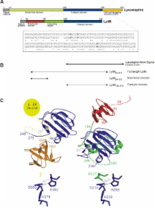
          Lysostaphin and the catalytic domain of LytM cleave pentaglycine crossbridges of Staphylococcus aureus peptidoglycan. The bacteriocin lysostaphin is secreted by Staphylococcus simulans biovar staphylolyticus and directed against the cell walls of competing S. aureus. LytM is produced by S. aureus as a latent autolysin and can be activated in vitro by the removal of an N-terminal domain and occluding region.

          Results

          We compared the efficacies of the lysostaphin and LytM catalytic domains using a newly developed model of chronic S. aureus infected eczema. Lysostaphin was effective, like in other models. In contrast, LytM was not significantly better than control. The different treatment outcomes could be correlated with in vitro properties of the proteins, including proteolytic stability, affinity to cell wall components other than peptidoglycan, and sensitivity to the ionic milieu.

          Conclusions

          Although lysostaphin and LytM cleave the same peptide bond in the peptidoglycan, the two enzymes have very different environmental requirements what is reflected in their contrasting performance in mouse eczema model.

          Related collections

          Most cited references38

          • Record: found
          • Abstract: found
          • Article: not found

          sigmaB modulates virulence determinant expression and stress resistance: characterization of a functional rsbU strain derived from Staphylococcus aureus 8325-4.

          The accessory sigma factor sigmaB controls a general stress response that is thought to be important for Staphylococcus aureus survival and may contribute to virulence. The strain of choice for genetic studies, 8325-4, carries a small deletion in rsbU, which encodes a positive regulator of sigmaB activity. Consequently, to enable the role of sigmaB in virulence to be addressed, we constructed an rsbU(+) derivative, SH1000, using a method that does not leave behind an antibiotic resistance marker. The phenotypic properties of SH1000 (8325-4 rsbU(+)) were characterized and compared to those of 8325-4, the rsbU mutant, parent strain. A recognition site for sigmaB was located in the promoter region of katA, the gene encoding the sole catalase of S. aureus, by primer extension analysis. However, catalase expression and activity were similar in SH1000 (8325-4 rsbU(+)), suggesting that this promoter may have a minor role in catalase expression under normal conditions. Restoration of sigmaB activity in SH1000 (8325-4 rsbU(+)) resulted in a marked decrease in the levels of the exoproteins SspA and Hla, and this is likely to be mediated by reduced expression of agr in this strain. By using Western blotting and a sarA-lacZ reporter assay, the levels of SarA were found to be similar in strains 8325-4 and SH1000 (8325-4 rsbU(+)) and sigB mutant derivatives of these strains. This finding contrasts with previous reports that suggested that SarA expression levels are altered when they are measured transcriptionally. Inactivation of sarA in each of these strains resulted in an expected decrease in agr expression; however, the relative level of agr in SH1000 (8325-4 rsbU(+)) remained less than the relative levels in 8325-4 and the sigB mutant derivatives. We suggest that SarA is not likely to be the effector in the overall sigmaB-mediated effect on agr expression.
            Bookmark
            • Record: found
            • Abstract: found
            • Article: not found

            MEROPS: the peptidase database

            Peptidases (proteolytic enzymes or proteases), their substrates and inhibitors are of great relevance to biology, medicine and biotechnology. The MEROPS database (http://merops.sanger.ac.uk) aims to fulfil the need for an integrated source of information about these. The organizational principle of the database is a hierarchical classification in which homologous sets of peptidases and protein inhibitors are grouped into protein species, which are grouped into families and in turn grouped into clans. Important additions to the database include newly written, concise text annotations for peptidase clans and the small molecule inhibitors that are outside the scope of the standard classification; displays to show peptidase specificity compiled from our collection of known substrate cleavages; tables of peptidase–inhibitor interactions; and dynamically generated alignments of representatives of each protein species at the family level. New ways to compare peptidase and inhibitor complements between any two organisms whose genomes have been completely sequenced, or between different strains or subspecies of the same organism, have been devised.
              Bookmark
              • Record: found
              • Abstract: found
              • Article: not found

              Lysostaphin disrupts Staphylococcus aureus and Staphylococcus epidermidis biofilms on artificial surfaces.

              Staphylococci often form biofilms, sessile communities of microcolonies encased in an extracellular matrix that adhere to biomedical implants or damaged tissue. Infections associated with biofilms are difficult to treat, and it is estimated that sessile bacteria in biofilms are 1,000 to 1,500 times more resistant to antibiotics than their planktonic counterparts. This antibiotic resistance of biofilms often leads to the failure of conventional antibiotic therapy and necessitates the removal of infected devices. Lysostaphin is a glycylglycine endopeptidase which specifically cleaves the pentaglycine cross bridges found in the staphylococcal peptidoglycan. Lysostaphin kills Staphylococcus aureus within minutes (MIC at which 90% of the strains are inhibited [MIC(90)], 0.001 to 0.064 microg/ml) and is also effective against Staphylococcus epidermidis at higher concentrations (MIC(90), 12.5 to 64 microg/ml). The activity of lysostaphin against staphylococci present in biofilms compared to those of other antibiotics was, however, never explored. Surprisingly, lysostaphin not only killed S. aureus in biofilms but also disrupted the extracellular matrix of S. aureus biofilms in vitro on plastic and glass surfaces at concentrations as low as 1 microg/ml. Scanning electron microscopy confirmed that lysostaphin eradicated both the sessile cells and the extracellular matrix of the biofilm. This disruption of S. aureus biofilms was specific for lysostaphin-sensitive S. aureus, as biofilms of lysostaphin-resistant S. aureus were not affected. High concentrations of oxacillin (400 microg/ml), vancomycin (800 microg/ml), and clindamycin (800 microg/ml) had no effect on the established S. aureus biofilms in this system, even after 24 h. Higher concentrations of lysostaphin also disrupted S. epidermidis biofilms.
                Bookmark

                Author and article information

                Journal
                BMC Microbiol
                BMC Microbiol
                BMC Microbiology
                BioMed Central
                1471-2180
                2012
                6 June 2012
                : 12
                : 97
                Affiliations
                [1 ]International Institute of Molecular and Cell Biology, Trojdena 4, 02-109, Warsaw, Poland
                [2 ]Max-Planck Institute of Molecular Cell Biology and Genetics, Pfotenhauerstr. 108, 01309, Dresden, Germany
                [3 ]Department of Rheumatology and Inflammation Research, University of Gothenburg, Guldhedsgatan 10A, S-413 46, Gothenburg, Sweden
                [4 ]Institute of Biochemistry and Biophysics, Polish Academy of Sciences, Pawinskiego 5a, 02-106, Warszawa, Poland
                Article
                1471-2180-12-97
                10.1186/1471-2180-12-97
                3413552
                22672475
                32feff84-d679-4a04-98bc-f448e321bcc6
                Copyright ©2012 Sabala et al.; licensee BioMed Central Ltd.

                This is an Open Access article distributed under the terms of the Creative Commons Attribution License ( http://creativecommons.org/licenses/by/2.0), which permits unrestricted use, distribution, and reproduction in any medium, provided the original work is properly cited.

                History
                : 5 January 2012
                : 6 June 2012
                Categories
                Research Article

                Microbiology & Virology
                Microbiology & Virology

                Comments

                Comment on this article