1
views
0
recommends
+1 Recommend
0 collections
    0
    shares
      • Record: found
      • Abstract: found
      • Article: found
      Is Open Access

      Presynaptic Rac1 controls synaptic strength through the regulation of synaptic vesicle priming

      research-article

      Read this article at

      Bookmark
          There is no author summary for this article yet. Authors can add summaries to their articles on ScienceOpen to make them more accessible to a non-specialist audience.

          Abstract

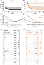
          Synapses contain a limited number of synaptic vesicles (SVs) that are released in response to action potentials (APs). Therefore, sustaining synaptic transmission over a wide range of AP firing rates and timescales depends on SV release and replenishment. Although actin dynamics impact synaptic transmission, how presynaptic regulators of actin signaling cascades control SV release and replenishment remains unresolved. Rac1, a Rho GTPase, regulates actin signaling cascades that control synaptogenesis, neuronal development, and postsynaptic function. However, the presynaptic role of Rac1 in regulating synaptic transmission is unclear. To unravel Rac1’s roles in controlling transmitter release, we performed selective presynaptic ablation of Rac1 at the mature mouse calyx of Held synapse. Loss of Rac1 increased synaptic strength, accelerated EPSC recovery after conditioning stimulus trains, and augmented spontaneous SV release with no change in presynaptic morphology or AZ ultrastructure. Analyses with constrained short-term plasticity models revealed faster SV priming kinetics and, depending on model assumptions, elevated SV release probability or higher abundance of tightly docked fusion-competent SVs in Rac1-deficient synapses. We conclude that presynaptic Rac1 is a key regulator of synaptic transmission and plasticity mainly by regulating the dynamics of SV priming and potentially SV release probability.

          Related collections

          Most cited references129

          • Record: found
          • Abstract: found
          • Article: not found

          Fiji: an open-source platform for biological-image analysis.

          Fiji is a distribution of the popular open-source software ImageJ focused on biological-image analysis. Fiji uses modern software engineering practices to combine powerful software libraries with a broad range of scripting languages to enable rapid prototyping of image-processing algorithms. Fiji facilitates the transformation of new algorithms into ImageJ plugins that can be shared with end users through an integrated update system. We propose Fiji as a platform for productive collaboration between computer science and biology research communities.
            Bookmark
            • Record: found
            • Abstract: found
            • Article: not found

            Multiple roles of calcium ions in the regulation of neurotransmitter release.

            The intracellular calcium concentration ([Ca(2+)]) has important roles in the triggering of neurotransmitter release and the regulation of short-term plasticity (STP). Transmitter release is initiated by quite high concentrations within microdomains, while short-term facilitation is strongly influenced by the global buildup of "residual calcium." A global rise in [Ca(2+)] also accelerates the recruitment of release-ready vesicles, thereby controlling the degree of short-term depression (STD) during sustained activity, as well as the recovery of the vesicle pool in periods of rest. We survey data that lead us to propose two distinct roles of [Ca(2+)] in vesicle recruitment: one accelerating "molecular priming" (vesicle docking and the buildup of a release machinery), the other promoting the tight coupling between releasable vesicles and Ca(2+) channels. Such coupling is essential for rendering vesicles sensitive to short [Ca(2+)] transients, generated during action potentials.
              Bookmark
              • Record: found
              • Abstract: found
              • Article: not found

              Ultrafast endocytosis at mouse hippocampal synapses

              To sustain neurotransmission, synaptic vesicles and their associated proteins must be recycled locally at synapses. Synaptic vesicles are thought to be regenerated ~20 s after fusion by the assembly of clathrin scaffolds or in ~1 s by the reversal of fusion pores via ‘kiss-and-run’ endocytosis. Here we use optogenetics to stimulate cultured hippocampal neurons with a single stimulus, rapidly freeze them after fixed intervals and examine the ultrastructure using electron microscopy – ‘flash-and-freeze’ electron microscopy. Docked vesicles fuse and collapse into the membrane within 30 ms of the stimulus. Compensatory endocytosis occurs with 50-100 ms at sites flanking the active zone. Invagination is blocked by inhibition of actin polymerization, and scission is blocked by inhibiting dynamin. Because intact synaptic vesicles are not recovered, this form of recycling is not compatible with kiss-and-run endocytosis; moreover it is 200-fold faster than clathrin-mediated endocytosis. It is likely that ‘ultrafast endocytosis’ is specialized to rapidly restore the surface area of the membrane.
                Bookmark

                Author and article information

                Contributors
                Role: Reviewing Editor
                Role: Senior Editor
                Journal
                eLife
                Elife
                eLife
                eLife
                eLife Sciences Publications, Ltd
                2050-084X
                10 October 2022
                2022
                : 11
                : e81505
                Affiliations
                [1 ] Department of Anatomy and Cell Biology, University of Iowa ( https://ror.org/036jqmy94) Iowa City United States
                [2 ] Department of Human Medicine, Carl-von-Ossietzky University Oldenburg ( https://ror.org/033n9gh91) Oldenburg Germany
                [3 ] Research Center Neurosensory Science, Carl-von-Ossietzky University Oldenburg ( https://ror.org/033n9gh91) Oldenburg Germany
                [4 ] Electron Microscopy Core Facility, Max Planck Florida Institute for Neuroscience ( https://ror.org/02rbfnr22) Jupiter United States
                [5 ] Department of Molecular Neurobiology, Max Planck Institute for Multidisciplinary Sciences ( https://ror.org/03av75f26) Göttingen Germany
                [6 ] Göttingen Graduate School for Neurosciences, Biophysics, and Molecular Biosciences Göttingen Germany
                [7 ] Department of Otolaryngology, Iowa Neuroscience Institute, University of Iowa ( https://ror.org/036jqmy94) Iowa City United States
                Stanford University School of Medicine ( https://ror.org/00f54p054) United States
                Stanford University School of Medicine ( https://ror.org/00f54p054) United States
                Stanford University School of Medicine ( https://ror.org/00f54p054) United States
                Stanford University School of Medicine ( https://ror.org/00f54p054) United States
                Max Planck Institute for Multidisciplinary Sciences ( https://ror.org/03av75f26) Germany
                Author information
                https://orcid.org/0000-0002-8953-2593
                https://orcid.org/0000-0003-3196-266X
                https://orcid.org/0000-0002-2825-9773
                https://orcid.org/0000-0003-0995-9667
                https://orcid.org/0000-0003-4310-3811
                https://orcid.org/0000-0003-3186-3231
                https://orcid.org/0000-0002-8926-5309
                https://orcid.org/0000-0002-7589-7612
                Article
                81505
                10.7554/eLife.81505
                9584605
                36214784
                34df2274-72cb-4320-bc6a-a7a9b186b60d
                © 2022, Keine et al

                This article is distributed under the terms of the Creative Commons Attribution License, which permits unrestricted use and redistribution provided that the original author and source are credited.

                History
                : 30 June 2022
                : 09 October 2022
                Funding
                Funded by: FundRef http://dx.doi.org/10.13039/100000055, National Institute on Deafness and Other Communication Disorders;
                Award ID: R01 DC014093
                Award Recipient :
                Funded by: FundRef http://dx.doi.org/10.13039/100000065, National Institute of Neurological Disorders and Stroke;
                Award ID: R01 NS110742
                Award Recipient :
                Funded by: FundRef http://dx.doi.org/10.13039/501100001659, Deutsche Forschungsgemeinschaft;
                Award ID: 420075000
                Award Recipient :
                Funded by: Max Planck Institute for Multidisciplinary Sciences;
                Award ID: open access funding
                Award Recipient :
                Funded by: Max Planck Florida Institute for Neuroscience;
                Award ID: open access funding
                Award Recipient :
                Funded by: FundRef http://dx.doi.org/10.13039/100008893, University of Iowa;
                Award ID: University Funds and University of Iowa Healthcare CCOM Distinguished Scholar Award
                Award Recipient :
                The funders had no role in study design, data collection and interpretation, or the decision to submit the work for publication.
                Categories
                Research Article
                Cell Biology
                Neuroscience
                Custom metadata
                A combination of genetic, electrophysiological, and modeling approaches reveals that presynaptic Rac1 is a key molecule that controls synaptic strength and plasticity by regulating the synaptic vesicle cycle steps controlling the number of fusion competent synaptic vesicles.

                Life sciences
                rac1,actin cytoskeleton,auditory brainstem,synaptic plasticity,synaptic transmission,synaptic vesicle replenishment,mouse

                Comments

                Comment on this article