18
views
0
recommends
+1 Recommend
0 collections
    0
    shares
      • Record: found
      • Abstract: found
      • Article: found
      Is Open Access

      Accuracy of Mobile Phone and Handheld Light Microscopy for the Diagnosis of Schistosomiasis and Intestinal Protozoa Infections in Côte d’Ivoire

      research-article

      Read this article at

      Bookmark
          There is no author summary for this article yet. Authors can add summaries to their articles on ScienceOpen to make them more accessible to a non-specialist audience.

          Abstract

          Background

          Handheld light microscopy using compact optics and mobile phones may improve the quality of health care in resource-constrained settings by enabling access to prompt and accurate diagnosis.

          Methodology

          Laboratory technicians were trained to operate two handheld diagnostic devices (Newton Nm1 microscope and a clip-on version of the mobile phone-based CellScope). The accuracy of these devices was compared to conventional light microscopy for the diagnosis of Schistosoma haematobium, S. mansoni, and intestinal protozoa infection in a community-based survey in rural Côte d’Ivoire. One slide of 10 ml filtered urine and a single Kato-Katz thick smear from 226 individuals were subjected to the Newton Nm1 microscope and CellScope for detection of Schistosoma eggs and compared to conventional microscopy. Additionally, 121 sodium acetate-acetic acid-formalin (SAF)-fixed stool samples were examined by the Newton Nm1 microscope and compared to conventional microscopy for the diagnosis of intestinal protozoa.

          Principal Findings

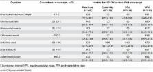
          The prevalence of S. haematobium, S. mansoni, Giardia intestinalis, and Entamoeba histolytica/E. dispar, as determined by conventional microscopy, was 39.8%, 5.3%, 20.7%, and 4.9%, respectively. The Newton Nm1 microscope had diagnostic sensitivities for S. mansoni and S. haematobium infection of 91.7% (95% confidence interval (CI) 59.8–99.6%) and 81.1% (95% CI 71.2–88.3%), respectively, and specificities of 99.5% (95% CI 97.0–100%) and 97.1% (95% CI 92.2–99.1%), respectively. The CellScope demonstrated sensitivities for S. mansoni and S. haematobium of 50.0% (95% CI 25.4–74.6%) and 35.6% (95% CI 25.9–46.4%), respectively, and specificities of 99.5% (95% CI 97.0–100%) and 100% (95% CI 86.7–100%), respectively. For G. intestinalis and E. histolytica/E. dispar, the Newton Nm1 microscope had sensitivity of 84.0% (95% CI 63.1–94.7%) and 83.3% (95% CI 36.5–99.1%), respectively, and 100% specificity.

          Conclusions/Significance

          Handheld diagnostic devices can be employed in community-based surveys in resource-constrained settings after minimal training of laboratory technicians to diagnose intestinal parasites.

          Author Summary

          Handheld light microscopes are new technologies that may be helpful in enabling better access to diagnostic testing for people living in resource-constrained settings in tropical and subtropical countries. Recent studies evaluating the accuracy of such devices have focused on their use by expert microscopists and were mainly conducted in laboratories. We evaluated the operating performance of two handheld microscopes (Newton Nm1 microscope and clip-on version of the reversed-lens CellScope) in comparison to conventional microscopy for the diagnosis of urogenital and intestinal schistosomiasis, when integrated into routine use in a community-based survey carried out in Côte d’Ivoire. Additionally, we evaluated the same microscopist’s diagnostic performance with the Newton Nm1 microscope for intestinal protozoa in a laboratory set-up. The Newton Nm1 microscope demonstrated excellent diagnostic sensitivity and specificity for schistosomiasis and intestinal protozoa. The CellScope had high specificity but only modest sensitivity for schistosomiasis diagnosis. Taken together, handheld diagnostic tools show promise to improve the quality of clinical and public health care delivered in resource-constrained settings.

          Related collections

          Most cited references22

          • Record: found
          • Abstract: found
          • Article: not found

          Amoebiasis.

          Amoebiasis is the second leading cause of death from parasitic disease worldwide. The causative protozoan parasite, Entamoeba histolytica, is a potent pathogen. Secreting proteinases that dissolve host tissues, killing host cells on contact, and engulfing red blood cells, E histolytica trophozoites invade the intestinal mucosa, causing amoebic colitis. In some cases amoebas breach the mucosal barrier and travel through the portal circulation to the liver, where they cause abscesses consisting of a few E histolytica trophozoites surrounding dead and dying hepatocytes and liquefied cellular debris. Amoebic liver abscesses grow inexorably and, at one time, were almost always fatal, but now even large abscesses can be cured by one dose of antibiotic. Evidence that what we thought was a single species based on morphology is, in fact, two genetically distinct species--now termed Entamoeba histolytica (the pathogen) and Entamoeba dispar (a commensal)--has turned conventional wisdom about the epidemiology and diagnosis of amoebiasis upside down. New models of disease have linked E histolytica induction of intestinal inflammation and hepatocyte programmed cell death to the pathogenesis of amoebic colitis and amoebic liver abscess.
            Bookmark
            • Record: found
            • Abstract: found
            • Article: not found

            Laboratory medicine in Africa: a barrier to effective health care.

            Providing health care in sub-Saharan Africa is a complex problem. Recent reports call for more resources to assist in the prevention and treatment of infectious diseases that affect this population, but policy makers, clinicians, and the public frequently fail to understand that diagnosis is essential to the prevention and treatment of disease. Access to reliable diagnostic testing is severely limited in this region, and misdiagnosis commonly occurs. Understandably, allocation of resources to diagnostic laboratory testing has not been a priority for resource-limited health care systems, but unreliable and inaccurate laboratory diagnostic testing leads to unnecessary expenditures in a region already plagued by resource shortages, promotes the perception that laboratory testing is unhelpful, and compromises patient care. We explore the barriers to implementing consistent testing within this region and illustrate the need for a more comprehensive approach to the diagnosis of infectious diseases, with an emphasis on making laboratory testing a higher priority.
              Bookmark
              • Record: found
              • Abstract: not found
              • Article: not found

              Schistosomiasis.

                Bookmark

                Author and article information

                Contributors
                Role: Editor
                Journal
                PLoS Negl Trop Dis
                PLoS Negl Trop Dis
                plos
                plosntds
                PLoS Neglected Tropical Diseases
                Public Library of Science (San Francisco, CA USA )
                1935-2727
                1935-2735
                27 June 2016
                June 2016
                : 10
                : 6
                : e0004768
                Affiliations
                [1 ]Unité de Formation et de Recherche Biosciences, Université Félix Houphouët-Boigny, Abidjan, Côte d’Ivoire
                [2 ]Centre Suisse de Recherches Scientifiques en Côte d’Ivoire, Abidjan, Côte d’Ivoire
                [3 ]Swiss Tropical and Public Health Institute, Basel, Switzerland
                [4 ]University of Basel, Basel, Switzerland
                [5 ]Department of Bioengineering, University of California–Berkeley, Berkeley, California, United States of America
                [6 ]Division of Infectious Diseases and Geographic Medicine, Stanford University School of Medicine, Stanford, California, United States of America
                [7 ]Divisions of Internal Medicine and Infectious Diseases, Toronto General Hospital, Toronto, Canada
                [8 ]Department of Medicine, University of Toronto, Toronto, Canada
                George Washington University, UNITED STATES
                Author notes

                The authors have declared that no competing interests exist.

                Conceived and designed the experiments: JC JRA IIB. Performed the experiments: JC MO IIB. Analyzed the data: JC JRA IIB. Contributed reagents/materials/analysis tools: JC MO MD DF JK JU EKN JRA IIB. Wrote the paper: JC MO MD DF JK JU EKN JRA IIB.

                Article
                PNTD-D-15-02005
                10.1371/journal.pntd.0004768
                4922625
                27348755
                354025f6-bddd-4374-92bd-de64c4f1e30c
                © 2016 Coulibaly et al

                This is an open access article distributed under the terms of the Creative Commons Attribution License, which permits unrestricted use, distribution, and reproduction in any medium, provided the original author and source are credited.

                History
                : 30 November 2015
                : 18 May 2016
                Page count
                Figures: 3, Tables: 2, Pages: 10
                Funding
                Funded by: Programme d'appui strategique a la recherche scientifique (PASRES) Cote d'Iovire
                Award ID: 33517764
                Award Recipient :
                Funded by: funder-id http://dx.doi.org/10.13039/501100004828, Grand Challenges Canada;
                Award ID: 0631-01-10
                Award Recipient :
                Funded by: MSH UHN AMO Innovation Fund
                Award Recipient :
                This study was funded by the Programme d’appui stratégique à la recherche scientifique (PASRES), Côte d’Ivoire (project no. 113) and Grand Challenges Canada 0631-01-10 ( www.grandchallenges.ca). In addition, IIB was supported by a grant from the MSH UHN AMO Innovation Fund. The funders played no role in the study design, data collection and analysis, decision to publish, or preparation of the manuscript.
                Categories
                Research Article
                Biology and Life Sciences
                Organisms
                Animals
                Invertebrates
                Helminths
                Schistosoma
                Schistosoma Haematobium
                Biology and Life Sciences
                Anatomy
                Digestive System
                Gastrointestinal Tract
                Medicine and Health Sciences
                Anatomy
                Digestive System
                Gastrointestinal Tract
                Biology and Life Sciences
                Organisms
                Protozoans
                Medicine and Health Sciences
                Diagnostic Medicine
                Biology and Life Sciences
                Organisms
                Animals
                Invertebrates
                Helminths
                Schistosoma
                Schistosoma Mansoni
                Research and Analysis Methods
                Microscopy
                Light Microscopy
                People and Places
                Population Groupings
                Professions
                Technicians
                Engineering and Technology
                Equipment
                Communication Equipment
                Cell Phones
                Custom metadata
                All relevant data are within the paper and its Supporting Information files.

                Infectious disease & Microbiology
                Infectious disease & Microbiology

                Comments

                Comment on this article