5
views
0
recommends
+1 Recommend
0 collections
    0
    shares
      • Record: found
      • Abstract: found
      • Article: found
      Is Open Access

      The EPO-FGF23 Signaling Pathway in Erythroid Progenitor Cells: Opening a New Area of Research

      review-article

      Read this article at

      Bookmark
          There is no author summary for this article yet. Authors can add summaries to their articles on ScienceOpen to make them more accessible to a non-specialist audience.

          Abstract

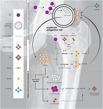
          We provide an overview of the evidence for an erythropoietin-fibroblast growth factor 23 (FGF23) signaling pathway directly influencing erythroid cells in the bone marrow. We outline its importance for red blood cell production, which might add, among others, to the understanding of bone marrow responses to endogenous erythropoietin in rare hereditary anemias. FGF23 is a hormone that is mainly known as the core regulator of phosphate and vitamin D metabolism and it has been recognized as an important regulator of bone mineralization. Osseous tissue has been regarded as the major source of FGF23. Interestingly, erythroid progenitor cells highly express FGF23 protein and carry the FGF receptor. This implies that erythroid progenitor cells could be a prime target in FGF23 biology. FGF23 is formed as an intact, biologically active protein (iFGF23) and proteolytic cleavage results in the formation of the presumed inactive C-terminal tail of FGF23 (cFGF23). FGF23-knockout or injection of an iFGF23 blocking peptide in mice results in increased erythropoiesis, reduced erythroid cell apoptosis and elevated renal and bone marrow erythropoietin mRNA expression with increased levels of circulating erythropoietin. By competitive inhibition, a relative increase in cFGF23 compared to iFGF23 results in reduced FGF23 receptor signaling and mimics the positive effects of FGF23-knockout or iFGF23 blocking peptide. Injection of recombinant erythropoietin increases FGF23 mRNA expression in the bone marrow with a concomitant increase in circulating FGF23 protein. However, erythropoietin also augments iFGF23 cleavage, thereby decreasing the iFGF23 to cFGF23 ratio. Therefore, the net result of erythropoietin is a reduction of iFGF23 to cFGF23 ratio, which inhibits the effects of iFGF23 on erythropoiesis and erythropoietin production. Elucidation of the EPO-FGF23 signaling pathway and its downstream signaling in hereditary anemias with chronic hemolysis or ineffective erythropoiesis adds to the understanding of the pathophysiology of these diseases and its complications; in addition, it provides promising new targets for treatment downstream of erythropoietin in the signaling cascade.

          Related collections

          Most cited references141

          • Record: found
          • Abstract: found
          • Article: not found

          Fibroblast growth factor 23 and risks of mortality and end-stage renal disease in patients with chronic kidney disease.

          A high level of the phosphate-regulating hormone fibroblast growth factor 23 (FGF-23) is associated with mortality in patients with end-stage renal disease, but little is known about its relationship with adverse outcomes in the much larger population of patients with earlier stages of chronic kidney disease. To evaluate FGF-23 as a risk factor for adverse outcomes in patients with chronic kidney disease. A prospective study of 3879 participants with chronic kidney disease stages 2 through 4 who enrolled in the Chronic Renal Insufficiency Cohort between June 2003 and September 2008. All-cause mortality and end-stage renal disease. At study enrollment, the mean (SD) estimated glomerular filtration rate (GFR) was 42.8 (13.5) mL/min/1.73 m(2), and the median FGF-23 level was 145.5 RU/mL (interquartile range [IQR], 96-239 reference unit [RU]/mL). During a median follow-up of 3.5 years (IQR, 2.5-4.4 years), 266 participants died (20.3/1000 person-years) and 410 reached end-stage renal disease (33.0/1000 person-years). In adjusted analyses, higher levels of FGF-23 were independently associated with a greater risk of death (hazard ratio [HR], per SD of natural log-transformed FGF-23, 1.5; 95% confidence interval [CI], 1.3-1.7). Mortality risk increased by quartile of FGF-23: the HR was 1.3 (95% CI, 0.8-2.2) for the second quartile, 2.0 (95% CI, 1.2-3.3) for the third quartile, and 3.0 (95% CI, 1.8-5.1) for the fourth quartile. Elevated fibroblast growth factor 23 was independently associated with significantly higher risk of end-stage renal disease among participants with an estimated GFR between 30 and 44 mL/min/1.73 m(2) (HR, 1.3 per SD of FGF-23 natural log-transformed FGF-23; 95% CI, 1.04-1.6) and 45 mL/min/1.73 m(2) or higher (HR, 1.7; 95% CI, 1.1-2.4), but not less than 30 mL/min/1.73 m(2). Elevated FGF-23 is an independent risk factor for end-stage renal disease in patients with relatively preserved kidney function and for mortality across the spectrum of chronic kidney disease.
            Bookmark
            • Record: found
            • Abstract: found
            • Article: not found

            Autosomal dominant hypophosphataemic rickets is associated with mutations in FGF23.

            (2000)
            Proper serum phosphate concentrations are maintained by a complex and poorly understood process. Identification of genes responsible for inherited disorders involving disturbances in phosphate homeostasis may provide insight into the pathways that regulate phosphate balance. Several hereditary disorders of isolated phosphate wasting have been described, including X-linked hypophosphataemic rickets (XLH), hypophosphataemic bone disease (HBD), hereditary hypophosphataemic rickets with hypercalciuria (HHRH) and autosomal dominant hypophosphataemic rickets (ADHR). Inactivating mutations of the gene PHEX, encoding a member of the neutral endopeptidase family of proteins, are responsible for XLH (refs 6,7). ADHR (MIM 193100) is characterized by low serum phosphorus concentrations, rickets, osteomalacia, lower extremity deformities, short stature, bone pain and dental abscesses. Here we describe a positional cloning approach used to identify the ADHR gene which included the annotation of 37 genes within 4 Mb of genomic sequence. We identified missense mutations in a gene encoding a new member of the fibroblast growth factor (FGF) family, FGF23. These mutations in patients with ADHR represent the first mutations found in a human FGF gene.
              Bookmark
              • Record: found
              • Abstract: found
              • Article: not found

              Hepcidin and iron regulation, 10 years later.

              Tomas Ganz (2011)
              Under evolutionary pressure to counter the toxicity of iron and to maintain adequate iron supply for hemoglobin synthesis and essential metabolic functions, humans and other vertebrates have effective mechanisms to conserve iron and to regulate its concentration, storage, and distribution in tissues. The iron-regulatory hormone hepcidin, first described 10 years ago, and its receptor and iron channel ferroportin control the dietary absorption, storage, and tissue distribution of iron. Hepcidin causes ferroportin internalization and degradation, thereby decreasing iron transfer into blood plasma from the duodenum, from macrophages involved in recycling senescent erythrocytes, and from iron-storing hepatocytes. Hepcidin is feedback regulated by iron concentrations in plasma and the liver and by erythropoietic demand for iron. Genetic malfunctions affecting the hepcidin-ferroportin axis are a main cause of iron overload disorders but can also cause iron-restricted anemias. Modulation of hepcidin and ferroportin expression during infection and inflammation couples iron metabolism to host defense and decreases iron availability to invading pathogens. This response also restricts the iron supply to erythropoietic precursors and may cause or contribute to the anemia associated with infections and inflammatory disorders.
                Bookmark

                Author and article information

                Contributors
                Journal
                Front Physiol
                Front Physiol
                Front. Physiol.
                Frontiers in Physiology
                Frontiers Media S.A.
                1664-042X
                26 March 2019
                2019
                : 10
                : 304
                Affiliations
                [1] 1Van Creveldkliniek, Department of Internal Medicine and Dermatology, University Medical Center Utrecht, Utrecht University , Utrecht, Netherlands
                [2] 2Department of Internal Medicine and Dermatology, University Medical Center Utrecht, Utrecht University , Utrecht, Netherlands
                [3] 3Department of Internal Medicine, Division of Nephrology, University Medical Center Groningen, University of Groningen , Groningen, Netherlands
                [4] 4Department of Clinical Chemistry and Haematology, University Medical Center Utrecht, Utrecht University , Utrecht, Netherlands
                Author notes

                Edited by: Lesley Jean Bruce, NHS Blood and Transplant, United Kingdom

                Reviewed by: Anna Rita Migliaccio, Icahn School of Medicine at Mount Sinai, United States; Angela Risso, University of Udine, Italy

                *Correspondence: Annelies J. van Vuren, A.J.vanVuren@ 123456umcutrecht.nl

                This article was submitted to Red Blood Cell Physiology, a section of the journal Frontiers in Physiology

                Article
                10.3389/fphys.2019.00304
                6443968
                30971944
                35b7d144-f670-4806-94d4-4e897666648f
                Copyright © 2019 van Vuren, Gaillard, Eisenga, van Wijk and van Beers.

                This is an open-access article distributed under the terms of the Creative Commons Attribution License (CC BY). The use, distribution or reproduction in other forums is permitted, provided the original author(s) and the copyright owner(s) are credited and that the original publication in this journal is cited, in accordance with accepted academic practice. No use, distribution or reproduction is permitted which does not comply with these terms.

                History
                : 10 December 2018
                : 07 March 2019
                Page count
                Figures: 2, Tables: 2, Equations: 0, References: 166, Pages: 16, Words: 0
                Categories
                Physiology
                Review

                Anatomy & Physiology
                fgf23,erythropoietin,anemia,osteoporosis,red blood cells
                Anatomy & Physiology
                fgf23, erythropoietin, anemia, osteoporosis, red blood cells

                Comments

                Comment on this article