10
views
0
recommends
+1 Recommend
0 collections
    0
    shares
      • Record: found
      • Abstract: found
      • Article: found
      Is Open Access

      Addison’s disease due to bilateral adrenal tuberculosis on 18F-fluorodeoxyglucose positron emission tomography computed tomography

      research-article
      Infectious Disease Reports
      PAGEPress Publications, Pavia, Italy
      Addison’s disease, tuberculosis, 18-FDG PET CT

      Read this article at

      Bookmark
          There is no author summary for this article yet. Authors can add summaries to their articles on ScienceOpen to make them more accessible to a non-specialist audience.

          Abstract

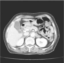
          We present a case of a 60-year-old woman diagnosed with disseminated tuberculosis with bilateral adrenalitis resulting in Addison’s disease. The 18-fluorodeoxyglucose (18-FDG) positron emission tomography (PET) computed tomography (CT) was performed, which revealed increased FDG uptake in the neck, mediastinal, and abdominal lymph nodes, and both adrenal glands, similar to the lesions noted on CT. We suspected the patient to have a lymphoma; therefore, axillary biopsy was performed, which revealed chronic granulomatous lesion with focal caseous necrosis.

          Related collections

          Most cited references10

          • Record: found
          • Abstract: not found
          • Article: not found

          Adrenal insufficiency.

          W Oelkers (1996)
            Bookmark
            • Record: found
            • Abstract: found
            • Article: not found

            Diagnosis of adrenal insufficiency.

            The cosyntropin stimulation test is the initial endocrine evaluation of suspected primary or secondary adrenal insufficiency. To critically review the utility of the cosyntropin stimulation test for evaluating adrenal insufficiency. The MEDLINE database was searched from 1966 to 2002 for all English-language papers related to the diagnosis of adrenal insufficiency. Studies with fewer than 5 persons with primary or secondary adrenal insufficiency or with fewer than 10 persons as normal controls were excluded. For secondary adrenal insufficiency, only studies that stratified participants by integrated tests of adrenal function were included. Summary receiver-operating characteristic (ROC) curves were generated from all studies that provided sensitivity and specificity data for 250-microg and 1-microg cosyntropin tests; these curves were then compared by using area under the curve (AUC) methods. All estimated values are given with 95% CIs. At a specificity of 95%, sensitivities were 97%, 57%, and 61% for summary ROC curves in tests for primary adrenal insufficiency (250-microg cosyntropin test), secondary adrenal insufficiency (250-microg cosyntropin test), and secondary adrenal insufficiency (1-microg cosyntropin test), respectively. The area under the curve for primary adrenal insufficiency was significantly greater than the AUC for secondary adrenal insufficiency for the high-dose cosyntropin test (P 0.5) for secondary adrenal insufficiency. At a specificity of 95%, summary ROC analysis for the 250-microg cosyntropin test yielded a positive likelihood ratio of 11.5 (95% CI, 8.7 to 14.2) and a negative likelihood ratio of 0.45 (CI, 0.30 to 0.60) for the diagnosis of secondary adrenal insufficiency. Cortisol response to cosyntropin varies considerably among healthy persons. The cosyntropin test performs well in patients with primary adrenal insufficiency, but the lower sensitivity in patients with secondary adrenal insufficiency necessitates use of tests involving stimulation of the hypothalamus if the pretest probability is sufficiently high. The operating characteristics of the 250-microg and 1-microg cosyntropin tests are similar.
              Bookmark
              • Record: found
              • Abstract: found
              • Article: not found

              18F-FDG PET in characterizing adrenal lesions detected on CT or MRI.

              The purpose of this study was to evaluate the ability of (18)F-FDG PET to characterize adrenal lesions in patients with proven or suspected cancers. A retrospective analysis was performed on 50 adrenal lesions in 41 patients, whose PET scans were done to evaluate the primary or metastatic disease. CT had shown 50 adrenal lesions in 41 patients and MRI had revealed 13 lesions in 10 patients. There were 34 patients with proven malignancy (28 lung cancer, 3 thyroid cancer, 2 colorectal cancer, and 1 lymphoma) and 7 with lung nodules. Of the 50 lesions, 18 were eventually determined to be malignant either by histopathology (n = 7) or by follow-up (n = 11). The remaining 32 lesions were proven or assumed to be benign by histopathology (n = 4) or clinical follow-up (n = 28). Unlike previously published reports, PET was interpreted as positive if the uptake was equal to or greater than that of the liver. No malignant lesion yielded a negative result on PET. Most lesions (13/18) showed significantly higher FDG uptake than that of the liver. In the remaining 5 lesions (2 metastases from neuroendocrine tumor, 2 early metastases, and 1 necrotic metastasis), FDG uptake was equal to or slightly higher than that of the liver. Of the 32 benign lesions, there were 2 lesions with uptake equal to or slightly higher than that of the liver, 3 with uptake less than the liver but more than the background, and 27 with uptake of the background. MRI identified 3 of the 13 lesions as false-positives but FDG PET correctly identified all 3 as benign. The other 10 adrenal lesions accurately diagnosed by MRI were also characterized by PET. FDG PET for characterization of adrenal lesions showed a sensitivity of 100%, a specificity of 94%, and an accuracy of 96%. FDG PET showed excellent diagnostic performance in differentiating adrenal lesions detected on CT or MRI. Because FDG PET has the additional advantage of evaluating the primary lesions as well as metastases, it could be cost-effective and the modality of choice for the characterization of adrenal lesions, especially in patients with malignancy.
                Bookmark

                Author and article information

                Journal
                Infect Dis Rep
                IDR
                Infectious Disease Reports
                PAGEPress Publications, Pavia, Italy
                2036-7430
                2036-7449
                25 September 2018
                05 September 2018
                : 10
                : 2
                : 7773
                Affiliations
                Department of Hematology-Oncology, College of Medicine, Yeungnam University , Daegu, Korea
                Author notes
                Department of Hematology-Oncology, College of Medicine, Yeungnam University, Daegu, Korea. +82.53.620.3826 - +82.53.654.8386. sakoh@ 123456yu.ac.kr

                Conflict of interest: the authors declare no potential conflict of interest.

                Article
                10.4081/idr.2018.7773
                6176475
                30344971
                35bfff48-5073-432c-8ca1-351036f1d853
                ©Copyright S.A.Koh, 2018

                This work is licensed under a Creative Commons Attribution NonCommercial 4.0 License (CC BY-NC 4.0).

                History
                : 05 June 2018
                : 23 June 2018
                : 25 June 2018
                Page count
                Figures: 3, Tables: 0, Equations: 0, References: 11, Pages: 2
                Categories
                Case Report

                addison’s disease,tuberculosis,18-fdg pet ct
                addison’s disease, tuberculosis, 18-fdg pet ct

                Comments

                Comment on this article