0
views
0
recommends
+1 Recommend
0 collections
    0
    shares
      • Record: found
      • Abstract: found
      • Article: found
      Is Open Access

      Spatial Transcriptomic Analysis of Pituitary Corticotroph Tumors Unveils Intratumor Heterogeneity

      Preprint
      research-article

      Read this article at

      Bookmark
          There is no author summary for this article yet. Authors can add summaries to their articles on ScienceOpen to make them more accessible to a non-specialist audience.

          Abstract

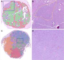
          Spatial transcriptomic (ST) analysis of tumors provides a novel approach on studying gene expression along with the localization of tumor cells in their environment to uncover spatial interactions. Herein, we present ST analysis of corticotroph pituitary neuroendocrine tumors (PitNETs) from formalin-fixed, paraffin-embedded (FFPE) tissues. We report that the in situ annotation of tumor tissue can be inferred from the gene expression profiles and is in concordance with the annotation made by a pathologist. Furthermore, relative gene expression in the tumor corresponds to common protein staining used in the evaluation of PitNETs, such as reticulin and Ki-67 index. Finally, we identify intratumor heterogeneity; clusters within the same tumor may present with different secretory capacity and transcriptomic profiles, unveiling potential intratumor cell variability with possible therapeutic interest. Together, our results provide the first attempt to clarify the spatial cell profile in PitNETs.

          Related collections

          Most cited references27

          • Record: found
          • Abstract: found
          • Article: not found

          Visualization and analysis of gene expression in tissue sections by spatial transcriptomics.

          Analysis of the pattern of proteins or messengerRNAs (mRNAs) in histological tissue sections is a cornerstone in biomedical research and diagnostics. This typically involves the visualization of a few proteins or expressed genes at a time. We have devised a strategy, which we call "spatial transcriptomics," that allows visualization and quantitative analysis of the transcriptome with spatial resolution in individual tissue sections. By positioning histological sections on arrayed reverse transcription primers with unique positional barcodes, we demonstrate high-quality RNA-sequencing data with maintained two-dimensional positional information from the mouse brain and human breast cancer. Spatial transcriptomics provides quantitative gene expression data and visualization of the distribution of mRNAs within tissue sections and enables novel types of bioinformatics analyses, valuable in research and diagnostics.
            Bookmark
            • Record: found
            • Abstract: found
            • Article: not found

            How does p53 induce apoptosis and how does this relate to p53-mediated tumour suppression?

            The tumour suppressor gene TP53 is mutated in ~50% of human cancers. In addition to its function in tumour suppression, p53 also plays a major role in the response of malignant as well as nontransformed cells to many anticancer therapeutics, particularly those that cause DNA damage. P53 forms a homotetrameric transcription factor that is reported to directly regulate ~500 target genes, thereby controlling a broad range of cellular processes, including cell cycle arrest, cell senescence, DNA repair, metabolic adaptation and cell death. For a long time, induction of apoptotic death in nascent neoplastic cells was regarded as the principal mechanism by which p53 prevents tumour development. This concept has, however, recently been challenged by the findings that in striking contrast to Trp53-deficient mice, gene-targeted mice that lack the critical effectors of p53-induced apoptosis do not develop tumours spontaneously. Remarkably, even mice lacking all mediators critical for p53-induced apoptosis, G1/S boundary cell cycle arrest and cell senescence do not develop any tumours spontaneously. In this review we discuss current understanding of the mechanisms by which p53 induces cell death and how this affects p53-mediated tumour suppression and the response of malignant cells to anticancer therapy.
              Bookmark
              • Record: found
              • Abstract: found
              • Article: not found

              The prevalence of pituitary adenomas: a systematic review.

              Pituitary adenomas display an array of hormonal and proliferative activity. Once primarily classified according to size (microadenomas, or = 1 cm), these tumors are now further classified according to immunohistochemistry and functional status. With these additional classifications in mind, the goals of the current study were to determine the prevalence of pituitary adenomas and to explore the clinical relevance of the findings. The authors conducted a metaanalysis of all existing English-language articles in MEDLINE. They used the search string (pituitary adenoma or pituitary tumor) and prevalence and selected relevant autopsy and imaging evaluation studies for inclusion. The authors found an overall estimated prevalence of pituitary adenomas of 16.7% (14.4% in autopsy studies and 22.5% in radiologic studies). Given the high frequency of pituitary adenomas and their potential for causing clinical pathologies, the findings of the current study suggest that early diagnosis and treatment of pituitary adenomas should have far-reaching benefits.
                Bookmark

                Author and article information

                Journal
                medRxiv
                MEDRXIV
                medRxiv
                Cold Spring Harbor Laboratory
                08 August 2023
                : 2023.08.04.23293576
                Affiliations
                [1 ]Section on Craniofacial Genetic Disorders, Eunice Kennedy Shriver National Institute of Child Health and Human Development, National Institutes of Health, Bethesda, Maryland, USA
                [2 ]Molecular Genomics Core, Eunice Kennedy Shriver National Institute of Child Health and Human Development, National Institutes of Health, Bethesda, Maryland, USA
                [3 ]Center for Immuno-Oncology, Center for Cancer Research, National Cancer Institute, National Institutes of Health, Bethesda, MD
                [4 ]Neurosurgery Unit for Pituitary and Inheritable Diseases, National Institute of Neurological Disorders and Stroke, National Institutes of Health, Bethesda, Maryland, USA
                [5 ]Laboratory of Pathology, Center for Cancer Research, National Institutes of Health, Bethesda, Maryland, USA
                [6 ]Unit on Hypothalamic and Pituitary Disorders, Eunice Kennedy Shriver National Institute of Child Health and Human Development, National Institutes of Health, Bethesda, Maryland, USA
                Author notes
                [*]

                Authors contributed equally

                Author contributions

                C.T. and F.R.F. designed the study; F.R.F., J.O.P., and C.P. performed experiments; C.T., C.S.F., F.R.F, and J.O.P. analyzed the data; C.T. and P.C. provided clinical care for the patients; C.T. wrote the manuscript with input from all authors.

                [** ] Corresponding author and person to whom reprints should be addressed: Christina Tatsi MD, MHSc, PhD, Eunice Kennedy Shriver National Institute of Child Health & Human Development (NICHD), 10 Center Drive, Building 10, Rom 1-3330, MSC1103, Bethesda, Maryland 20892, christina.tatsi3@ 123456nih.gov
                Author information
                http://orcid.org/0000-0002-2882-4585
                Article
                10.1101/2023.08.04.23293576
                10473795
                37662403
                35d051a1-0e6c-4e9c-9640-96f5053a9f9b

                This work is licensed under a Creative Commons Attribution-NonCommercial-NoDerivatives 4.0 International License, which allows reusers to copy and distribute the material in any medium or format in unadapted form only, for noncommercial purposes only, and only so long as attribution is given to the creator.

                History
                Funding
                Funded by: Intramural Research Program, Eunice Kennedy Shriver National Institute of Child Health & Human Development (NICHD), National Institutes of Health
                Categories
                Article

                transcriptomics,cushing disease,pituitary neuroendocrine tumor

                Comments

                Comment on this article