12
views
0
recommends
+1 Recommend
0 collections
    0
    shares
      • Record: found
      • Abstract: found
      • Article: found
      Is Open Access

      Accuracy of shoulder ultrasound examination for diagnosis of rotator cuff pathologies: a single-center retrospective study

      research-article

      Read this article at

      Bookmark
          There is no author summary for this article yet. Authors can add summaries to their articles on ScienceOpen to make them more accessible to a non-specialist audience.

          ABSTRACT

          BACKGROUND:

          Shoulder pathologies need accurate diagnosis for best management and treatment provided to patients.

          OBJECTIVE:

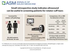
          Determine the diagnostic sensitivity, specificity and accuracy of shoulder ultrasonography (US).

          DESIGN:

          Retrospective, analytical.

          SETTING:

          Tertiary care center in Riyadh, Saudi Arabia.

          PATIENTS AND METHODS:

          We included all shoulder exams performed between January 2010 and December 2016 that met the inclusion criteria. Data was collected retrospectively from the a picture archiving and communication system and patient records. The patients were evaluated using US for the presence of rotator cuff tears and classified into intact, full-thickness tear, partial-thickness tear, tendinosis, subacromial/subdeltoid bursitis and acromioclavicular joint degenerative changes. The US findings were correlated with the shoulder MRI study findings. The time interval between the US examination and MRI ranged from 0 to 180 days (6 months).

          MAIN OUTCOME MEASURES:

          To compare the sensitivity, specificity and accuracy of shoulder US studies in the detection of rotator cuff pathologies in comparison to MRI findings.

          SAMPLE SIZE AND CHARACTERISTICS:

          86 (60 females, 26 male), mean age 53.7 years (range, 19-85).

          RESULTS:

          The sensitivity, specificity and accuracy of US for the detection of full-thickness supraspinatus tears compared with those of MRI were 86%, 82% and 83%, respectively. The sensitivity, specificity and accuracy of US for the detection of partial-thickness supraspinatus tears compared with those of MRI were 38%, 70% and 58%, respectively. Overall PPV, NPV, sensitivity, specificity and accuracy of US for the detection of full-thickness tears compared with those of MRI were 35%, 97%, 78%, 83% and 83%, respectively. For partial-thickness tears, the overall PPV, NPV, sensitivity, specificity and accuracy of US compared with those of MRI were 51%, 60%, 51%, 60% and 56%, respectively.

          CONCLUSION:

          Overall, US has high sensitivity, specificity and accuracy for the detection of full-thickness tears compared with the detection of partial-thickness tears.

          LIMITATIONS:

          Small sample size and retrospective.

          CONFLICT OF INTEREST:

          None.

          Abstract

          Related collections

          Most cited references18

          • Record: found
          • Abstract: found
          • Article: not found

          Prevalence and risk factors of a rotator cuff tear in the general population.

          Little information is available about the epidemiology of rotator cuff tears in a population-based study. The purpose of this study was to elucidate the true prevalence of rotator cuff tears regardless of the presence or absence of symptoms in the general population and to assess the relationship between tears and their backgrounds. A medical check-up was conducted for residents of a mountain village in Japan. The subjects consisted of 683 people (total of 1,366 shoulders), including 229 males and 454 females with a mean age of 57.9 years (range, 22-87). We examined their background factors, physical examinations and ultrasonographic examinations on both shoulders. Rotator cuff tears were present in 20.7% and the prevalence increased with age. Thirty-six percent of the subjects with current symptoms had rotator cuff tears, while 16.9% of the subjects without symptoms also had rotator cuff tears. Rotator cuff tears in the general population were most commonly associated with elderly patients, males, affected the dominant arm, engaged in heavy labor, having a history of trauma, positive for impingement sign, showed lesser active forward elevation and weaker muscle strength in abduction and external rotation. A logistic regression analysis revealed the risk factors for a rotator cuff tear to be a history of trauma, dominant arm and age. 20.7% of 1,366 shoulders had full-thickness rotator cuff tears in the general population. The risk factors for rotator cuff tear included a history of trauma, dominant arm and age. Level 3.
            Bookmark
            • Record: found
            • Abstract: found
            • Article: not found

            Epidemiology, natural history, and indications for treatment of rotator cuff tears.

            The etiology of rotator cuff disease is likely multifactorial, including age-related degeneration and microtrauma and macrotrauma. The incidence of rotator cuff tears increases with aging with more than half of individuals in their 80s having a rotator cuff tear. Smoking, hypercholesterolemia, and genetics have all been shown to influence the development of rotator cuff tearing. Substantial full-thickness rotator cuff tears, in general, progress and enlarge with time. Pain, or worsening pain, usually signals tear progression in both asymptomatic and symptomatic tears and should warrant further investigation if the tear is treated conservatively. Larger (>1-1.5 cm) symptomatic full-thickness cuff tears have a high rate of tear progression and, therefore, should be considered for earlier surgical repair in younger patients if the tear is reparable and there is limited muscle degeneration to avoid irreversible changes to the cuff, including tear enlargement and degenerative muscle changes. Smaller symptomatic full-thickness tears have been shown to have a slower rate of progression, similar to partial-thickness tears, and can be considered for initial nonoperative treatment due to the limited risk for rapid tear progression. In both small full-thickness tears and partial-thickness tears, increasing pain should alert physicians to obtain further imaging as it can signal tear progression. Natural history data, along with information on factors affecting healing after rotator cuff repair, can help guide surgeons in making appropriate decisions regarding the treatment of rotator cuff tears. The management of rotator cuff tears should be considered in the context of the risks and benefits of operative versus nonoperative treatment. Tear size and acuity, the presence of irreparable changes to the rotator cuff or glenohumeral joint, and patient age should all be considered in making this decision. Initial nonoperative care can be safely undertaken in older patients (>70 years old) with chronic tears; in patients with irreparable rotator cuff tears with irreversible changes, including significant atrophy and fatty infiltration, humeral head migration, and arthritis; in patients of any age with small ( 1 cm-1.5 cm) acute tears or young patients with full-thickness tears who have a significant risk for the development of irreparable rotator cuff changes.
              Bookmark
              • Record: found
              • Abstract: found
              • Article: not found

              Static and Dynamic Shoulder Imaging to Predict Initial Effectiveness and Recurrence After Ultrasound-Guided Subacromial Corticosteroid Injections.

              To explore factors contributing to initial effectiveness and recurrence after ultrasound (US)-guided subacromial corticosteroid injections by assessing clinical measurements and static and dynamic shoulder US images.
                Bookmark

                Author and article information

                Journal
                Ann Saudi Med
                Ann Saudi Med
                asm
                Annals of Saudi Medicine
                King Faisal Specialist Hospital and Research Centre
                0256-4947
                0975-4466
                May-June 2019
                30 May 2019
                : 39
                : 3
                : 162-171 (pp. 162-171)
                Affiliations
                [1]From the Department of Radiology, King Faisal Specialist Hospital and Research Centre, Riyadh, Saudi Arabia
                Author notes
                Correspondence: Rafat Saeed Mohtasib · Department of Biomedical Physics, King Faisal Specialist Hospital and Research Centre, PO Box 3354, Riyadh 11211 Saudi Arabia · T: 966-500-944-447 · rmohtasib@ 123456kfshrc.edu.sa · ORCID: https://orcid.org/0000-0002-9041-2177
                Author information
                https://orcid.org/0000-0002-9041-2177
                Article
                10.5144_0256-4947.2019.162
                10.5144/0256-4947.2019.162
                6832332
                31215221
                363a3791-8d22-4424-907b-d9c3c2a5f054
                Copyright © 2019, Annals of Saudi Medicine, Saudi Arabia

                This is an open access article under the Creative Commons Attribution-NonCommercial-NoDerivatives 4.0 International License (CC BY-NC-ND). The details of which can be accessed at http://creativecommons.org/licenses/by-nc-nd/4.0/

                History
                : 13 October 2018
                : 05 March 2019
                Funding
                None.
                Categories
                Original Article

                Medicine
                Medicine

                Comments

                Comment on this article