4
views
0
recommends
+1 Recommend
0 collections
    0
    shares
      • Record: found
      • Abstract: found
      • Article: found
      Is Open Access

      New Insights Into the Role and Mechanism of Partial Epithelial-Mesenchymal Transition in Kidney Fibrosis

      review-article

      Read this article at

      Bookmark
          There is no author summary for this article yet. Authors can add summaries to their articles on ScienceOpen to make them more accessible to a non-specialist audience.

          Abstract

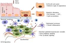
          Epithelial-mesenchymal transition (EMT) is described as the process in which injured renal tubular epithelial cells undergo a phenotype change, acquiring mesenchymal characteristics and morphing into fibroblasts. Initially, it was widely thought of as a critical mechanism of fibrogenesis underlying chronic kidney disease. However, evidence that renal tubular epithelial cells can cross the basement membrane and become fibroblasts in the renal interstitium is rare, leading to debate about the existence of EMT. Recent research has demonstrated that after injury, renal tubular epithelial cells acquire mesenchymal characteristics and the ability to produce a variety of profibrotic factors and cytokines, but remain attached to the basement membrane. On this basis, a new concept of “partial epithelial-mesenchymal transition (pEMT)” was proposed to explain the contribution of renal epithelial cells to renal fibrogenesis. In this review, we discuss the concept of pEMT and the most recent findings related to this process, including cell cycle arrest, metabolic alternation of epithelial cells, infiltration of immune cells, epigenetic regulation as well as the novel signaling pathways that mediate this disturbed epithelial-mesenchymal communication. A deeper understanding of the role and the mechanism of pEMT may help in developing novel therapies to prevent and halt fibrosis in kidney disease.

          Related collections

          Most cited references87

          • Record: found
          • Abstract: found
          • Article: not found

          Origin and function of myofibroblasts in kidney fibrosis.

          Myofibroblasts are associated with organ fibrosis, but their precise origin and functional role remain unknown. We used multiple genetically engineered mice to track, fate map and ablate cells to determine the source and function of myofibroblasts in kidney fibrosis. Through this comprehensive analysis, we identified that the total pool of myofibroblasts is split, with 50% arising from local resident fibroblasts through proliferation. The nonproliferating myofibroblasts derive through differentiation from bone marrow (35%), the endothelial-to-mesenchymal transition program (10%) and the epithelial-to-mesenchymal transition program (5%). Specific deletion of Tgfbr2 in α-smooth muscle actin (αSMA)(+) cells revealed the importance of this pathway in the recruitment of myofibroblasts through differentiation. Using genetic mouse models and a fate-mapping strategy, we determined that vascular pericytes probably do not contribute to the emergence of myofibroblasts or fibrosis. Our data suggest that targeting diverse pathways is required to substantially inhibit the composite accumulation of myofibroblasts in kidney fibrosis.
            Bookmark
            • Record: found
            • Abstract: found
            • Article: not found

            Mechanisms of tubulointerstitial fibrosis.

            The pathologic paradigm for renal progression is advancing tubulointerstitial fibrosis. Whereas mechanisms underlying fibrogenesis have grown in scope and understanding in recent decades, effective human treatment to directly halt or even reverse fibrosis remains elusive. Here, we examine key features mediating the molecular and cellular basis of tubulointerstitial fibrosis and highlight new insights that may lead to novel therapies. How to prevent chronic kidney disease from progressing to renal failure awaits even deeper biochemical understanding.
              Bookmark
              • Record: found
              • Abstract: found
              • Article: not found

              Renal tubule injury: a driving force toward chronic kidney disease.

              Renal tubules are the major component of the kidney and are vulnerable to a variety of injuries including hypoxia, proteinuria, toxins, metabolic disorders, and senescence. It has long been believed that tubules are the victim of injury. In this review, we shift this concept to renal tubules as a driving force in the progression of kidney diseases. In response to injury, tubular epithelial cells undergo changes and function as inflammatory and fibrogenic cells, with the consequent production of various bioactive molecules that drive interstitial inflammation and fibrosis. Innate immune-sensing receptors on the tubular epithelium also aggravate immune responses. Necroinflammation, an autoamplification loop between tubular cell death and interstitial inflammation, leads to the exacerbation of renal injury. Furthermore, tubular cells also play an active role in progressive renal injury via emerging mechanisms associated with a partial epithelial-mesenchymal transition, cell-cycle arrest at both G1/S and G2/M check points, and metabolic disorder. Thus, a better understanding the mechanisms by which tubular injury drives inflammation and fibrosis is necessary for the development of therapeutics to halt the progression of chronic kidney disease.
                Bookmark

                Author and article information

                Contributors
                Journal
                Front Physiol
                Front. Physiol.
                Frontiers in Physiology
                Frontiers Media S.A.
                1664-042X
                15 September 2020
                2020
                : 11
                : 569322
                Affiliations
                [1] 1Department of Nephrology, Shanghai East Hospital, Tongji University School of Medicine , Shanghai, China
                [2] 2Department of Medicine, Rhode Island Hospital and Alpert Medical School, Brown University , Providence, RI, United States
                Author notes

                Edited by: Guiomar Nascimento Gomes, Federal University of São Paulo, Brazil

                Reviewed by: Youhua Liu, University of Pittsburgh, United States; Danilo Candido De Almeida, Federal University of São Paulo, Brazil

                *Correspondence: Shougang Zhuang, shougang_zhuang@ 123456brown.edu

                This article was submitted to Renal and Epithelial Physiology, a section of the journal Frontiers in Physiology

                Article
                10.3389/fphys.2020.569322
                7522479
                33041867
                363ad1f7-b21b-48af-bd86-203c82b8756f
                Copyright © 2020 Sheng and Zhuang.

                This is an open-access article distributed under the terms of the Creative Commons Attribution License (CC BY). The use, distribution or reproduction in other forums is permitted, provided the original author(s) and the copyright owner(s) are credited and that the original publication in this journal is cited, in accordance with accepted academic practice. No use, distribution or reproduction is permitted which does not comply with these terms.

                History
                : 03 June 2020
                : 26 August 2020
                Page count
                Figures: 1, Tables: 1, Equations: 0, References: 111, Pages: 11, Words: 0
                Funding
                Funded by: National Natural Science Foundation of China 10.13039/501100001809
                Funded by: National Institutes of Health 10.13039/100000002
                Categories
                Physiology
                Review

                Anatomy & Physiology
                renal fibrosis,myofibroblast,epithelial-mesenchymal transition,partial epithelial-mesenchymal transition,chronic kidney disease

                Comments

                Comment on this article