14
views
0
recommends
+1 Recommend
0 collections
    0
    shares
      • Record: found
      • Abstract: found
      • Article: found
      Is Open Access

      α2AP mediated myofibroblast formation and the development of renal fibrosis in unilateral ureteral obstruction

      research-article

      Read this article at

      Bookmark
          There is no author summary for this article yet. Authors can add summaries to their articles on ScienceOpen to make them more accessible to a non-specialist audience.

          Abstract

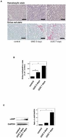
          Renal fibrosis is the final common pathway of a wide variety of chronic kidney diseases. Myofibroblast formation via the differentiation of from tissue-resident fibroblasts and bone marrow-derived mesenchymal stem cells (MSCs), and epithelial-to-mesenchymal transition (EMT) is known to play a pivotal role in the development of renal fibrosis. However, the detailed mechanisms underlying this disorder remain unclear. We herein investigated the role of alpha 2-antiplasmin (α2AP) in myofibroblast formation and the development of renal fibrosis. We observed the development of renal fibrosis using unilateral ureteral obstruction (UUO). α2AP had accumulated in the UUO-induced obstructed kidneys and α2AP deficiency attenuated UUO-induced renal fibrosis in mice. The degree of myofibroblast formation in the obstructed kidneys of α2AP −/− mice was less than that in α2AP +/+ mice. In vitro, α2AP induced myofibroblast formation in renal tubular epithelial cells (RTECs), renal fibrosblasts, and bone marrow-derived mesenchymal stem cells (MSCs). α2AP also induced the production of TGF-β, which is known to be a key regulator of myofibroblast formation and fibrosis. α2AP-induced the TGF-β production was significantly reduced by SP600125, c-Jun N-terminal kinase (JNK) specific inhibitor. Our findings suggest that α2AP induces myofibroblast formation in the obstructed kidneys, and mediates the development of renal fibrosis.

          Related collections

          Most cited references33

          • Record: found
          • Abstract: found
          • Article: not found

          Origin and function of myofibroblasts in kidney fibrosis.

          Myofibroblasts are associated with organ fibrosis, but their precise origin and functional role remain unknown. We used multiple genetically engineered mice to track, fate map and ablate cells to determine the source and function of myofibroblasts in kidney fibrosis. Through this comprehensive analysis, we identified that the total pool of myofibroblasts is split, with 50% arising from local resident fibroblasts through proliferation. The nonproliferating myofibroblasts derive through differentiation from bone marrow (35%), the endothelial-to-mesenchymal transition program (10%) and the epithelial-to-mesenchymal transition program (5%). Specific deletion of Tgfbr2 in α-smooth muscle actin (αSMA)(+) cells revealed the importance of this pathway in the recruitment of myofibroblasts through differentiation. Using genetic mouse models and a fate-mapping strategy, we determined that vascular pericytes probably do not contribute to the emergence of myofibroblasts or fibrosis. Our data suggest that targeting diverse pathways is required to substantially inhibit the composite accumulation of myofibroblasts in kidney fibrosis.
            Bookmark
            • Record: found
            • Abstract: found
            • Article: not found

            Making sense of latent TGFbeta activation.

            TGFbeta is secreted as part of a latent complex that is targeted to the extracellular matrix. A variety of molecules, 'TGFbeta activators,' release TGFbeta from its latent state. The unusual temporal discontinuity of TGFbeta synthesis and action and the panoply of TGFbeta effects contribute to the interest in TGF-beta. However, the logical connections between TGFbeta synthesis, storage and action are obscure. We consider the latent TGFbeta complex as an extracellular sensor in which the TGFbeta propeptide functions as the detector, latent-TGFbeta-binding protein (LTBP) functions as the localizer, and TGF-beta functions as the effector. Such a view provides a logical continuity for various aspects of TGFbeta biology and allows us to appreciate TGFbeta biology from a new perspective.
              Bookmark
              • Record: found
              • Abstract: found
              • Article: not found

              Targeted disruption of TGF-beta1/Smad3 signaling protects against renal tubulointerstitial fibrosis induced by unilateral ureteral obstruction.

              Tubulointerstitial fibrosis is the final common result of a variety of progressive injuries leading to chronic renal failure. Transforming growth factor-beta (TGF-beta) is reportedly upregulated in response to injurious stimuli such as unilateral ureteral obstruction (UUO), causing renal fibrosis associated with epithelial-mesenchymal transition (EMT) of the renal tubules and synthesis of extracellular matrix. We now show that mice lacking Smad3 (Smad3ex8/ex8), a key signaling intermediate downstream of the TGF-beta receptors, are protected against tubulointerstitial fibrosis following UUO as evidenced by blocking of EMT and abrogation of monocyte influx and collagen accumulation. Culture of primary renal tubular epithelial cells from wild-type or Smad3-null mice confirms that the Smad3 pathway is essential for TGF-beta1-induced EMT and autoinduction of TGF-beta1. Moreover, mechanical stretch of the cultured epithelial cells, mimicking renal tubular distention due to accumulation of urine after UUO, induces EMT following Smad3-mediated upregulation of TGF-beta1. Exogenous bone marrow monocytes accelerate EMT of the cultured epithelial cells and renal tubules in the obstructed kidney after UUO dependent on Smad3 signaling. Together the data demonstrate that the Smad3 pathway is central to the pathogenesis of interstitial fibrosis and suggest that inhibitors of this pathway may have clinical application in the treatment of obstructive nephropathy.
                Bookmark

                Author and article information

                Journal
                Sci Rep
                Sci Rep
                Scientific Reports
                Nature Publishing Group
                2045-2322
                06 August 2014
                2014
                : 4
                : 5967
                Affiliations
                [1 ]Department of Clinical Pathological Biochemistry, Faculty of Pharmaceutical Science , Doshisha Women's Collage of Liberal Arts, 97-1 Kodo Kyo-tanabe, Kyoto, 610-0395, Japan
                [2 ]Department of Physiology II. Kinki University School of Medicine , Osaka-sayama, Japan
                [3 ]Department of Dermatology, Gifu University Graduate School of Medicine , Yanagido Gifu, Japan
                [4 ]Department of Food Science and Nutrition, Kinki University School of Agriculture , Nara, Japan
                Author notes
                Article
                srep05967
                10.1038/srep05967
                5380014
                25095732
                3718b1e7-f3d6-43ac-b73c-2ae3e22101ec
                Copyright © 2014, Macmillan Publishers Limited. All rights reserved

                This work is licensed under a Creative Commons Attribution 4.0 International License. The images or other third party material in this article are included in the article's Creative Commons license, unless indicated otherwise in the credit line; if the material is not included under the Creative Commons license, users will need to obtain permission from the license holder in order to reproduce the material. To view a copy of this license, visit http://creativecommons.org/licenses/by/4.0/

                History
                : 16 January 2014
                : 15 July 2014
                Categories
                Article

                Uncategorized
                Uncategorized

                Comments

                Comment on this article