8
views
0
recommends
+1 Recommend
0 collections
    0
    shares
      • Record: found
      • Abstract: found
      • Article: found
      Is Open Access

      Susceptibility weighted imaging: Clinical applications and future directions

      review-article

      Read this article at

      Bookmark
          There is no author summary for this article yet. Authors can add summaries to their articles on ScienceOpen to make them more accessible to a non-specialist audience.

          Abstract

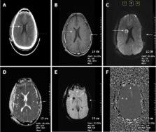
          Susceptibility weighted imaging (SWI) is a recently developed magnetic resonance imaging (MRI) technique that is increasingly being used to narrow the differential diagnosis of many neurologic disorders. It exploits the magnetic susceptibility differences of various compounds including deoxygenated blood, blood products, iron and calcium, thus enabling a new source of contrast in MR. In this review, we illustrate its basic clinical applications in neuroimaging. SWI is based on a fully velocity-compensated, high-resolution, three dimensional gradient-echo sequence using magnitude and phase images either separately or in combination with each other, in order to characterize brain tissue. SWI is particularly useful in the setting of trauma and acute neurologic presentations suggestive of stroke, but can also characterize occult low-flow vascular malformations, cerebral microbleeds, intracranial calcifications, neurodegenerative diseases and brain tumors. Furthermore, advanced MRI post-processing technique with quantitative susceptibility mapping, enables detailed anatomical differentiation based on quantification of brain iron from SWI raw data.

          Related collections

          Most cited references69

          • Record: found
          • Abstract: found
          • Article: not found

          Susceptibility weighted imaging (SWI).

          Susceptibility differences between tissues can be utilized as a new type of contrast in MRI that is different from spin density, T1-, or T2-weighted imaging. Signals from substances with different magnetic susceptibilities compared to their neighboring tissue will become out of phase with these tissues at sufficiently long echo times (TEs). Thus, phase imaging offers a means of enhancing contrast in MRI. Specifically, the phase images themselves can provide excellent contrast between gray matter (GM) and white matter (WM), iron-laden tissues, venous blood vessels, and other tissues with susceptibilities that are different from the background tissue. Also, for the first time, projection phase images are shown to demonstrate tissue (vessel) continuity. In this work, the best approach for combining magnitude and phase images is discussed. The phase images are high-pass-filtered and then transformed to a special phase mask that varies in amplitude between zero and unity. This mask is multiplied a few times into the original magnitude image to create enhanced contrast between tissues with different susceptibilities. For this reason, this method is referred to as susceptibility-weighted imaging (SWI). Mathematical arguments are presented to determine the number of phase mask multiplications that should take place. Examples are given for enhancing GM/WM contrast and water/fat contrast, identifying brain iron, and visualizing veins in the brain. Copyright 2004 Wiley-Liss, Inc.
            Bookmark
            • Record: found
            • Abstract: found
            • Article: not found

            Imaging iron stores in the brain using magnetic resonance imaging.

            For the last century, there has been great physiological interest in brain iron and its role in brain function and disease. It is well known that iron accumulates in the brain for people with Huntington's disease, Parkinson's disease, Alzheimer's disease, multiple sclerosis, chronic hemorrhage, cerebral infarction, anemia, thalassemia, hemochromatosis, Hallervorden-Spatz, Down syndrome, AIDS and in the eye for people with macular degeneration. Measuring the amount of nonheme iron in the body may well lead to not only a better understanding of the disease progression but an ability to predict outcome. As there are many forms of iron in the brain, separating them and quantifying each type have been a major challenge. In this review, we present our understanding of attempts to measure brain iron and the potential of doing so with magnetic resonance imaging. Specifically, we examine the response of the magnetic resonance visible iron in tissue that produces signal changes in both magnitude and phase images. These images seem to correlate with brain iron content, perhaps ferritin specifically, but still have not been successfully exploited to accurately and precisely quantify brain iron. For future quantitative studies of iron content we propose four methods: correlating R2' and phase to iron content; applying a special filter to the phase to obtain a susceptibility map; using complex analysis to extract the product of susceptibility and volume content of the susceptibility source; and using early and late echo information to separately predict susceptibility and volume content.
              Bookmark
              • Record: found
              • Abstract: found
              • Article: not found

              Susceptibility-weighted imaging: technical aspects and clinical applications, part 2.

              Susceptibility-weighted imaging (SWI) has continued to develop into a powerful clinical tool to visualize venous structures and iron in the brain and to study diverse pathologic conditions. SWI offers a unique contrast, different from spin attenuation, T1, T2, and T2* (see Susceptibility-Weighted Imaging: Technical Aspects and Clinical Applications, Part 1). In this clinical review (Part 2), we present a variety of neurovascular and neurodegenerative disease applications for SWI, covering trauma, stroke, cerebral amyloid angiopathy, venous anomalies, multiple sclerosis, and tumors. We conclude that SWI often offers complementary information valuable in the diagnosis and potential treatment of patients with neurologic disorders.
                Bookmark

                Author and article information

                Contributors
                Journal
                World J Radiol
                WJR
                World Journal of Radiology
                Baishideng Publishing Group Inc
                1949-8470
                28 April 2018
                28 April 2018
                : 10
                : 4
                : 30-45
                Affiliations
                Department of Radiology, Sisli Hamidiye Etfal Training and Research Hospital, University of Health Sciences, Istanbul 34371, Turkey
                Division of Neuroradiology, Department of Radiology, Johns Hopkins Medical Institution, Baltimore, MI 21287, United States
                Author notes

                Author contributions: All authors equally contributed to this paper with conception and design of the study, literature review and analysis, drafting, critical revision and editing, and final approval of the final version.

                Correspondence to: Ahmet Mesrur Halefoglu, MD, Professor, Department of Radiology, Sisli Hamidiye Etfal Training and Research Hospital, University of Health Sciences, Birlik sok, Parksaray ap, No: 17/4, Istanbul 34371, Turkey. halefoglu@ 123456hotmail.com

                Telephone: +90-212-3735000 Fax: +90-212-2415015

                Article
                jWJR.v10.i4.pg30
                10.4329/wjr.v10.i4.30
                5971274
                29849962
                3737379a-903e-4d18-abf8-6f0764d8ea6b
                ©The Author(s) 2018. Published by Baishideng Publishing Group Inc. All rights reserved.

                This article is an open-access article which was selected by an in-house editor and fully peer-reviewed by external reviewers. It is distributed in accordance with the Creative Commons Attribution Non Commercial (CC BY-NC 4.0) license, which permits others to distribute, remix, adapt, build upon this work non-commercially, and license their derivative works on different terms, provided the original work is properly cited and the use is non-commercial.

                History
                : 17 March 2018
                : 8 April 2018
                : 20 April 2018
                Categories
                Review

                quantitative susceptibility mapping,brain,ischemia,magnetic resonance imaging,susceptibility weighted imaging

                Comments

                Comment on this article