
- Record: found
- Abstract: found
- Article: found
Spinal microcircuits comprising dI3 interneurons are necessary for motor functional recovery following spinal cord transection
Read this article at
Abstract
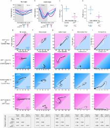
The spinal cord has the capacity to coordinate motor activities such as locomotion. Following spinal transection, functional activity can be regained, to a degree, following motor training. To identify microcircuits involved in this recovery, we studied a population of mouse spinal interneurons known to receive direct afferent inputs and project to intermediate and ventral regions of the spinal cord. We demonstrate that while dI3 interneurons are not necessary for normal locomotor activity, locomotor circuits rhythmically inhibit them and dI3 interneurons can activate these circuits. Removing dI3 interneurons from spinal microcircuits by eliminating their synaptic transmission left locomotion more or less unchanged, but abolished functional recovery, indicating that dI3 interneurons are a necessary cellular substrate for motor system plasticity following transection. We suggest that dI3 interneurons compare inputs from locomotor circuits with sensory afferent inputs to compute sensory prediction errors that then modify locomotor circuits to effect motor recovery.
eLife digest
Circuits of nerve cells, or neurons, in the spinal cord control movement. After an injury to the spinal cord, the connections between the brain and spinal neurons may be severed, meaning that the spinal circuits can no longer work properly. This loss of communication between the brain and the spinal cord often leads to paralysis below the level of the injury.
There are currently no effective treatments for individuals who have lost the ability to walk following spinal cord injury. However, the spinal cord retains circuits that are sufficient to restore walking and these circuits can be activated with training. That is, rehabilitative training can lead to improvements in movement by promoting spinal cord plasticity – the ability of other neurons in the spinal cord to take over the roles of the severed neurons. By understanding how rehabilitation leads to these improvements following injury, new strategies could be developed to optimize the recovery process.
Previous research showed that spinal neurons called dI3 interneurons are involved in short term adjustments of movement. Could these interneurons also be involved in longer term adaptations?
Bui, Stifani et al. compared normal mice with genetically engineered mice that had dI3 interneurons “removed” from their circuits. This revealed that although dI3 interneurons in mice are integrated with spinal circuits that are involved in walking, they are not necessary for normal walking. Following the severing of the spinal cord, the experimental mice, unlike the normal mice, did not recover the ability to step. Thus, circuits comprising dI3 interneurons are necessary for recovering the ability to move after an injury.
Now that Bui, Stifani et al. have identified this essential circuit, the next step is to investigate how dI3 interneurons promote spinal cord plasticity. Understanding these mechanisms could help to develop therapies that enhance rehabilitation-assisted improvement of movement following spinal cord injury.
Related collections
Most cited references56
- Record: found
- Abstract: found
- Article: not found
NIH Image to ImageJ: 25 years of image analysis.
- Record: found
- Abstract: found
- Article: not found